


Total list price:
Log in to view your online price 
Select your location.
*e-commerce not available for this region.


The total SMAD3 kit detects cellular SMAD3 and can be used as a normalization assay with our phospho-SMAD3 kit to enable optimal investigation of TGF-ß signaling.
| Feature | Specification |
|---|---|
| Application | Cell Signaling |
| Sample Volume | 16 µL |
The total SMAD3 kit detects cellular SMAD3 and can be used as a normalization assay with our phospho-SMAD3 kit to enable optimal investigation of TGF-ß signaling.
The total SMAD3 cellular assay kit is designed to monitor the expression level of SMAD3, whether phosphorylated or unphosphorylated. It is compatible with our Phospho-SMAD3 kit, and enables analysis of phosphorylated and total proteins from a single sample for better readout of TGF-ß signaling activity.
| Application |
Cell Signaling
|
|---|---|
| Brand |
HTRF
|
| Detection Modality |
HTRF
|
| Lysis Buffer Compatibility |
Lysis Buffer 1
Lysis Buffer 3
Lysis Buffer 4
Lysis Buffer 5
|
| Molecular Modification |
Total
|
| Product Group |
Kit
|
| Sample Volume |
16 µL
|
| Shipping Conditions |
Shipped in Dry Ice
|
| Target Class |
Phosphoproteins
|
| Target Species |
Human
|
| Technology |
TR-FRET
|
| Therapeutic Area |
Cardiovascular
Metabolism/Diabetes
NASH/Fibrosis
Oncology & Inflammation
|
| Unit Size |
500 assay points
|
The Total-SMAD3 assay quantifies the expression level of SMAD3 in a cell lysate. Contrary to Western Blot, the assay is entirely plate-based and does not require gels, electrophoresis or transfer. The Total-SMAD3 assay uses two labeled antibodies: one coupled to a donor fluorophore, the other to an acceptor. Both antibodies are highly specific for a distinct epitope on the protein. In presence of SMAD3 in a cell extract, the addition of these conjugates brings the donor fluorophore into close proximity with the acceptor and thereby generates a FRET signal. Its intensity is directly proportional to the concentration of the protein present in the sample, and provides a means of assessing the proteins expression under a no-wash assay format.
The 2 plate protocol involves culturing cells in a 96-well plate before lysis then transferring lysates to a 384-well low volume detection plate before adding Total-SMAD3 HTRF detection reagents. This protocol enables the cells' viability and confluence to be monitored.

Detection of total SMAD3 with HTRF reagents can be performed in a single plate used for culturing, stimulation and lysis. No washing steps are required. This HTS designed protocol enables miniaturization while maintaining robust HTRF quality.

Human LX-2* and HepG2 lines were plated (50,000 and 200,000 cells/well respectively) and cultured overnight in complete culture medium, 37°C - 5% CO2. Cells were then treated with increasing concentrations of TGF-ß1 for 30 minutes at 37°C - 5% CO2. After medium removal, cells were lysed with 50 µL of supplemented lysis buffer #4 for 30 minutes at RT under gentle shaking, and 16 µL of lysate were transferred twice over into a low volume white microplate before adding 4 µL of the HTRF phospho-SMAD3 or total SMAD3 detection antibodies. The HTRF signal was recorded after an overnight incubation. In both lines, TGF-ß1 treatment induces SMAD3 phosphorylation on Ser423/425, while the expression level of the protein is not significantly affected.
*LX-2 cell line provided by EMD Millipore (Part #SCC064)


Mouse NIH/3T3 and C2C12 lines were plated (100,000 cells/well) and cultured 2H in complete culture medium, 37°C - 5% CO2. The day after, the cells were treated with increasing concentrations of TGF-ß1 for 30 minutes at 37°C - 5% CO2. After medium removal, the cells were lysed with 50 µL of supplemented lysis buffer #4 for 30 minutes at RT under gentle shaking, and 16 µL of lysate were transferred twice over into a low volume white microplate before adding 4 µL of the HTRF phospho-SMAD3 or total SMAD3 detection antibodies. The HTRF signal was recorded after an overnight incubation. In both lines, TGF-ß1 promotes the activation of SMAD3 by phosphorylation on Ser423/425, whereas the expression level of the protein remains fairly stable.


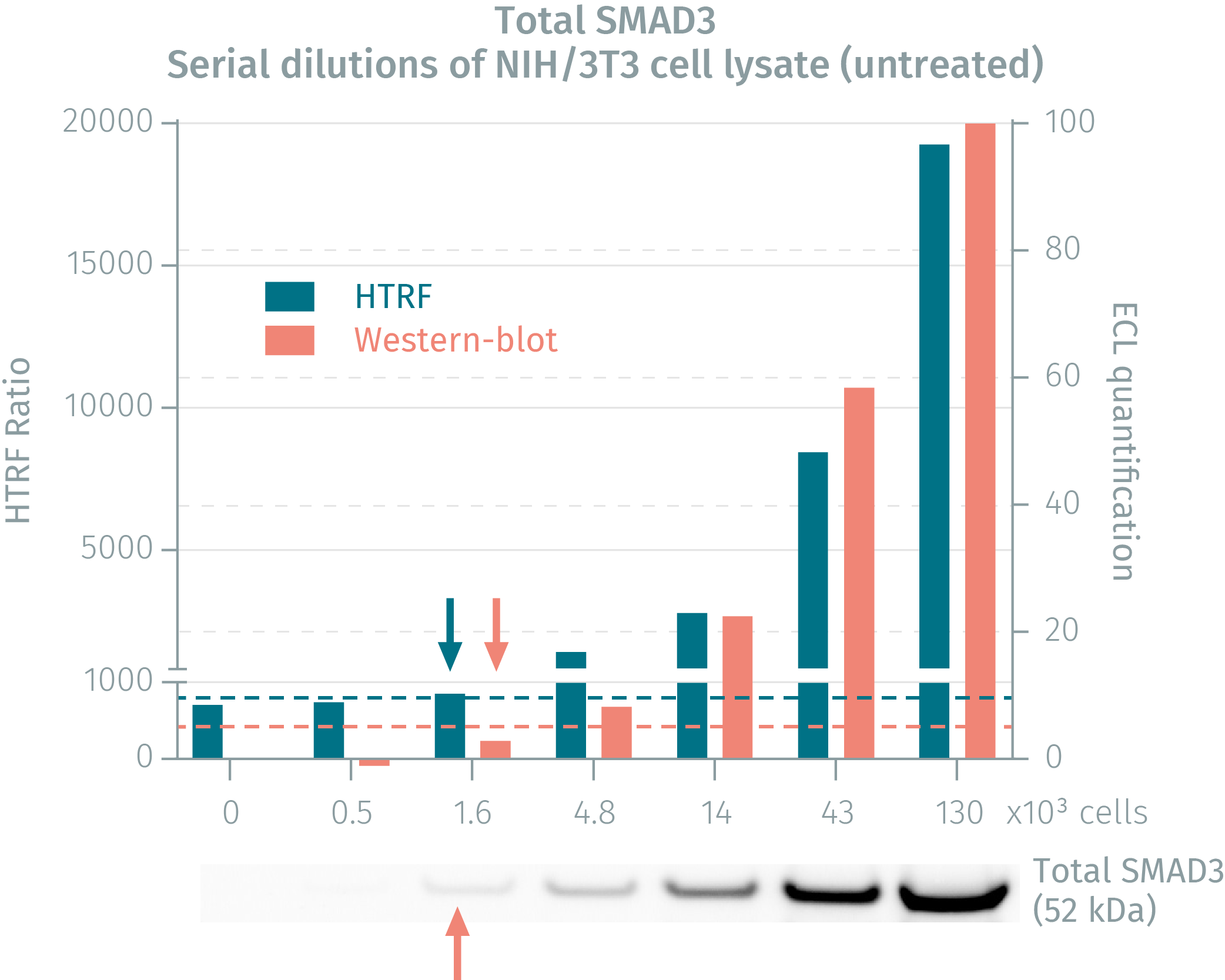
Cells were cultured to 80% confluency and treated with TGF-ß1 for 30 minutes. Following lysis, soluble supernatants were collected via centrifugation. Serial dilutions of ysates were performed and transferred into a low volume white microplate before finally adding HTRF phospho-SMAD3 detection reagents. Equal amounts of lysates were used for a side by side comparison between HTRF and Western Blot. The results reveal that the HTRF phospho-SMAD3 assay is 16-fold more sensitive than the Western Blot.
TGF-ß signaling is mediated by complexes of TßRI and TßRII which activate the intracellular SMAD3 by phosphorylation. The binding of the TGF-ß ligand on TßRII triggers the recruitment of TßRI into the ligand-receptor complex. TßRII autophosphorylates then transphosphorylates TßRI. Activated TßRI in turn phosphorylates SMAD3 on Ser423 and Ser425, enabling its oligomerization with SMAD4. This complex then translocates to the nucleus and acts as a transcription factor with coactivators and corepressors to regulate the expression of multiple genes involved in cell growth, apoptosis, proliferation, migration, and differentiation, as well as in extracellular matrix remodeling and immune/inflammatory responses. Inhibitory SMAD6 and SMAD7 are involved in feedback inhibition of the pathway.

Are you looking for resources, click on the resource type to explore further.
Discover the benefits of using HTRF assays to characterize liver fibrosis.
Several molecular markers have been investigated as...
Discover the versatility and precision of Homogeneous Time-Resolved Fluorescence (HTRF) technology. Our HTRF portfolio offers a...
This guide provides you an overview of HTRF applications in several therapeutic areas.
Loading...


We are here to answer your questions.


