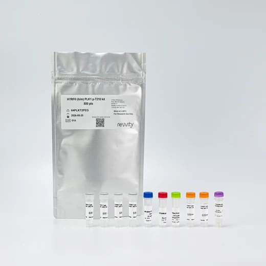

HTRF Human and Mouse Phospho-PLK1 (Thr210) Detection Kit, 500 Assay Points


HTRF Human and Mouse Phospho-PLK1 (Thr210) Detection Kit, 500 Assay Points


| Feature | Specification |
|---|---|
| Application | Cell Signaling |
| Sample Volume | 16 µL |


Product information
Overview
This HTRF cell-based assay enables the rapid, quantitative detection of PLK1 phosphorylated at Threonine 210. Polo-like kinase 1 (PLK1) is the principle member of the well conserved serine/threonine kinase family and is essential for cell division (mitosis). As such, it is a key regulator of the G2/M checkpoint of the cell cycle
PLK1 activation for cell cycle regulation: PLK1 is activated by phosphorylation on Thr210 by AurA kinase. Activated PLK1 phosphorylates CDC25.
PLK1 is a critical protein responsible for mitosis and cell cycle progression/cell proliferation. This makes it an attractive cancer treatment target.
How it works
Phospho-PLK1 (Thr210) assay principle
The HTRF Phospho-PLK1 (Thr210) assay measures PLK1 when phosphorylated at Thr210. Unlike Western Blot, the assay is entirely plate-based and does not require gels, electrophoresis, or transfer. The Phospho-PLK1 (Thr210) assay uses 2 labeled antibodies: one with a donor fluorophore, the other with an acceptor. The first antibody was selected for its specific binding to the phosphorylated motif on the protein, and the second for its ability to recognize the protein independently of its phosphorylation state. Protein phosphorylation enables an immune-complex formation involving the two labeled antibodies and which brings the donor fluorophore into close proximity to the acceptor, thereby generating a FRET signal. Its intensity is directly proportional to the concentration of phosphorylated protein present in the sample, and provides a means of assessing the protein’s phosphorylation state under a no-wash assay format.

Phospho-PLK1 (Thr210 ) two-plate assay protocol
The two-plate protocol involves culturing cells in a 96-well plate before lysis, then transferring lysates into a 384-well low volume detection plate before the addition of the Phospho-PLK1 (Thr210) HTRF detection reagents. This protocol enables the cells' viability and confluence to be monitored.

Phospho-PLK1 (Thr210 ) one-plate assay protocol
Detection of Total PLK1 with HTRF reagents can be performed in a single plate used for culturing, stimulation, and lysis. No washing steps are required.
This HTS-designed protocol enables miniaturization while maintaining robust HTRF quality.

Assay validation
Activation of Total and Phospho PLK1 Thr210 measured on HeLa cells
HeLa cells were plated at 20,000 cells/ well in a 96-well culture-treated plate in complete culture medium, and incubated overnight at 37°C, 5% CO2. Cells were then treated with increasing concentrations of Nocodazole for 16h at 37°C, 5% CO2. They were next lysed with 50 µl of supplemented lysis buffer #1 (1X) for 30 min at RT under gentle shaking.
After cell lysis, 16 µL of lysate were transferred into a 384-well low volume white microplate and 4 µL of the HTRF Total or Phospho PLK1 Thr210 detection reagents were added. The HTRF signals were recorded after 18h of incubation at room temperature.
As expected, Nocodazole induces the increase expression and phosphorylation of PLK1 with a dose-dependent manner with equivalent EC50 60-70 nM.

Inhibitor characterization with Phospho-PLK1 (Thr210) and Total PLK1 on HeLa cells
HeLa cells that express PLK1 were plated at 20,000 cells /well in 96-well culture-treated plates in complete culture medium, and incubated overnight at 37°C, 5% CO2. Cells were then co-treated with 200 nM of Nocozazole and increasing concentrations of the PLK1 activator Doxorubicin, 2 AuroraA inhibitors MLN8054 & Alisertib, and the PROTAC compound CC885 for 16h at 37°C, 5% CO2. Next, the medium was removed, and the cells were lysed with 50 µl of supplemented lysis buffer #1 (1X) for 30 min at RT under gentle shaking.
After cell lysis, 16 µL of lysate were transferred into a 384-well low volume white microplate and 4 µL of the HTRF Phospho PLK1 Thr210 and Total-PLK1 detection reagents were added. The HTRF signals were recorded after an overnight incubation at room temperature.
As expected, all of these compounds induced a loss of PLK1 phosphorylation, with different EC50: 54 nM for Doxorubicin, around 6 µM for MLN8054, 28 nM for Alisertib, and 68 nM for the PROTAC CC885.
Interestingly, the expression of PLK1 decreased in the same way for all the compounds except for MLN8054.


Validation on various Human and Mouse cell lines
Adherent human & mouse cells HeLa, MCF7, Hep-G2, and NIH 3T3 were seeded at 25,000 cells / well in a 96-well microplate. After a 24H incubation, the cells were treated with 300 nM of nocodazole for 16h at 37°C. The cells were then lysed with supplemented lysis buffer #1, and 16 µL of lysate were transferred into a 384-well low volume white microplate before the addition of 4 µL of the HTRF Phospho PLK1 Thr210 detection reagents. The HTRF signals were recorded after an overnight incubation.
The HTRF Phospho PLK1 Thr210 assay efficiently detected Phosphorylated PLK1 in various cellular models expressing different levels of the protein.

HTRF Phospho PLK1 (Thr210) assay compared to Western Blot
HeLa cells were cultured in a T175 flask in a complete culture medium for 24h at 37°C, 5% CO2. The cells were treated with 300 nM of Nocodazole compound for 16h at 37°C, 5% CO2. They were then lysed with 3 mL of supplemented lysis buffer #1 (1x) for 30 minutes at RT under gentle shaking.
Serial dilutions of the cell lysate were performed using supplemented lysis buffer #1 (1x), and 16 µL of each dilution were transferred into a low volume white microplate before the addition of 4 µL of HTRF Phospho Thr210 PLK1 detection reagents.
Equal amounts of lysates were used for a side-by-side comparison between HTRF and Western Blot.
In these conditions, the HTRF Phospho PLK1 assay was 32 times more sensitive than the Western Blot technique.

Simplified pathway
PLK1 signaling pathway
PLK1 for Polo-like kinase 1, has a key role in the progression of mitosis and recent evidence suggest its important involvement in regulating the G2/M checkpoint, in DNA damage and replication stress response, and in cell death pathways. Signaling mechanism is quite complex and involves several positive (Bora/AuroraA pathway) and negative feedback loops (CHK1/ATR or CHK2/ATM pathways). PLK1 activation for cell cycle regulation: PLK1 is activated by phosphorylation on Thr210 by AurA kinase. Then Activated PLK1 phosphorylates CDC25. In Autophagy/Apoptosis, PLK1 has been implicated in various cell death processes via various signaling pathways incluing mTOR, cmyc and p53. Recently, new roles of PLK1 have been reported in literature on its implication in the regulation of inflammation and immunological responses, including NFkB and RIG-I/MAV signaling. All these biological processes are altered in tumors and, considering that PLK1 is often found overexpressed in several tumor types; this overexpression is associated with poor patient prognosis. Consequently, its targeting has emerged as a promising anti-cancer therapeutic strategy.

Specifications
| Application |
Cell Signaling
|
|---|---|
| Brand |
HTRF
|
| Detection Modality |
HTRF
|
| Lysis Buffer Compatibility |
Lysis Buffer 1
Lysis Buffer 2
Lysis Buffer 4
|
| Molecular Modification |
Phosphorylation
|
| Product Group |
Kit
|
| Sample Volume |
16 µL
|
| Shipping Conditions |
Shipped in Dry Ice
|
| Target Class |
Phosphoproteins
|
| Target Species |
Human
Mouse
|
| Technology |
TR-FRET
|
| Unit Size |
500 assay points
|
Video gallery
Resources
Are you looking for resources, click on the resource type to explore further.
Discover the versatility and precision of Homogeneous Time-Resolved Fluorescence (HTRF) technology. Our HTRF portfolio offers a...
This guide provides you an overview of HTRF applications in several therapeutic areas.
SDS, COAs, manuals and more
Are you looking for technical documents related to the product? We have categorized them in dedicated sections below. Explore now or request your COA/TDS, SDS, or IFU/manual.
- LanguageEnglishCountryUnited States
- LanguageFrenchCountryFrance
- LanguageGermanCountryGermany
- Lot Number01ALot DateApril 3, 2026
- Resource TypeManualLanguageEnglishCountry-


How can we help you?
We are here to answer your questions.


