


Total list price:
Log in to view your online price 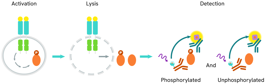
Select your location.
*e-commerce not available for this region.


This HTRF kit allows for the cell-based quantitative detection of total ARID1A.
10% OFF lab essentials when you buy on Revvity.com or through your Revvity punchout with promo code.
Promo ends 5/21. Terms and conditions apply.
| Feature | Specification |
|---|---|
| Application | Cell Signaling |
| Sample Volume | 16 µL |
This HTRF kit allows for the cell-based quantitative detection of total ARID1A.
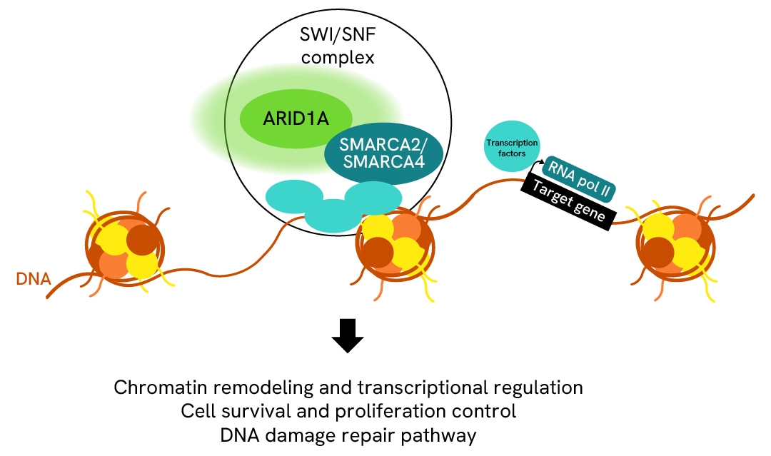
ARID1A (AT-rich interactive domain-containing protein 1A) is a member of the SWI/SNF (SWItch/Sucrose Non-Fermenting) complex and is involved in transcriptional activation and repression of selected genes by chromatin remodeling. ARID1A plays a crucial role in mammalian development and acts as a tumor suppressor, regulating gene transcription, participating in DNA damage response, and influencing tumor immune microenvironment and signaling pathways.
| Application |
Cell Signaling
|
|---|---|
| Brand |
HTRF
|
| Detection Modality |
HTRF
|
| Lysis Buffer Compatibility |
Lysis Buffer 4
|
| Molecular Modification |
Total
|
| Product Group |
Kit
|
| Sample Volume |
16 µL
|
| Shipping Conditions |
Shipped in Dry Ice
|
| Target |
ARID1A
|
| Target Class |
Phosphoproteins
|
| Target Species |
Human
Mouse
|
| Technology |
TR-FRET
|
| Therapeutic Area |
Oncology
|
| Unit Size |
10,000 assay points
|
The Total ARID1A assay measures ARID1A levels in cells. Unlike Western Blot, the assay is entirely plate-based and does not require gels, electrophoresis, or transfer. The assay uses 2 antibodies, one labeled with a donor fluorophore and the other with an acceptor. Both antibodies are highly specific for a distinct epitope on the protein.
In presence of ARID1A this enables an immune-complex formation involving both labeled antibodies, and which brings the donor fluorophore into close proximity to the acceptor, thereby generating a FRET signal.
Its intensity is directly proportional to the concentration of total protein present in the sample and provides a means of assessing the protein's phosphorylation state under a no-wash assay format.
The two-plate protocol involves culturing cells in a 96-well plate before lysis, then transferring lysates into a 384-well low volume detection plate before the addition of Total ARID1A HTRF detection reagents. This protocol allows for the cells' viability and confluence to be monitored.

Detection of Total ARID1A with HTRF reagents can be performed in a single plate used for culturing, stimulation, and lysis. No washing steps are required. This HTS designed protocol allows miniaturization while maintaining robust HTRF quality.

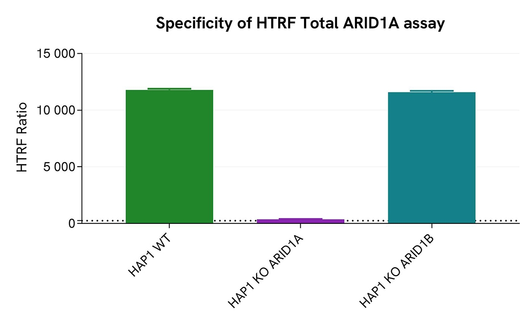
HAP1 WT, KO ARID1A, and KO ARID1B cells were plated in a 96-well plate (50,000 cells/well) in complete culture medium, and incubated overnight at 37°C, 5% CO2. After culture medium removal, cells were lysed with 50 µl of supplemented Lysis Buffer #4 for 30 min at room temperature under gentle shaking. After cell lysis, 16 µL of cell lysate were transferred into a HTRF 384-well low volume white microplate, and then then 4 µL of the HTRF Total ARID1A detection reagents were added. The HTRF signal was recorded using an EnVision Nexus reader after an overnight-incubation at room temperature.
As depicted in the figure, HTRF signals were detected in the HAP1 WT and KO ARID1B cell lines. Conversely, near-background signal was observed in the KO ARID1A cell line, demonstrating the specificity of the HTRF Total ARID1A assay for the detection of the ARID1A protein.
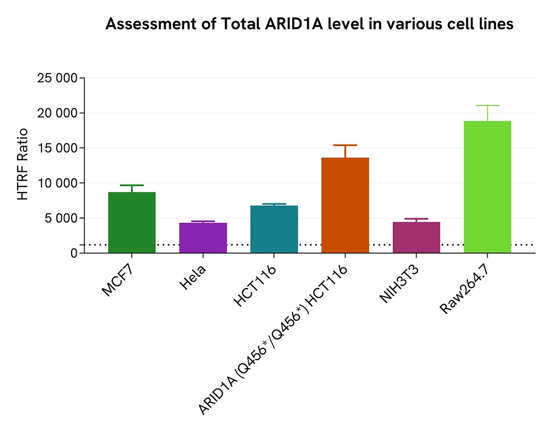
HeLa, MCF7, HCT116, ARID1A (Q456*/Q456*) HCT116, NIH3T3, and Raw264.7 cells were cultured in flasks in complete culture medium, and incubated 48 h at 37°C, 5% CO2. After culture medium removal, cells were lysed with 3 mL of supplemented Lysis Buffer #4 for 30 min at room temperature under gentle shaking. After cell lysis, 16 µL of cell lysate (dilution 1/8) were transferred into a HTRF 384-well low volume white microplate, then 4 µL of the HTRF Total RNA Pol II detection reagents were added. The HTRF signal was recorded using an HTRF compatible reader after an overnight-incubation at room temperature.
The HTRF Total ARID1A assay demonstrated its ability to detect Total ARID1A in various cellular models expressing different levels of the protein.
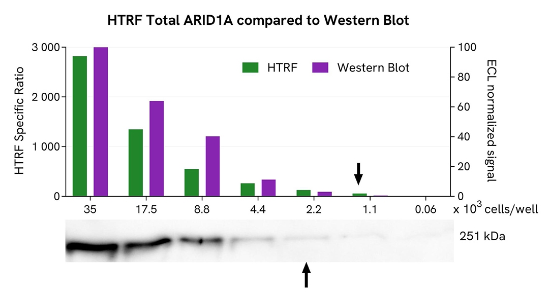
HeLa cells were cultured in flasks and incubated 48 h at 37°C, 5% CO2. After culture medium removal, cells were lysed with 3 mL of supplemented Lysis Buffer #4 for 30 min at room temperature under gentle shaking. Equal amounts of cell lysates (13 µL) were used for a side-by-side comparison of Western Blot and HTRF techniques.
Using the HTRF Total ARID1A assay, 1,100 cells/well were enough to detect a significant signal, while more than 2,200 cells were needed to obtain a minimal chemiluminescent signal using Western Blot. Therefore, in these conditions, the HTRF Total ARID1A assay is 2 times more sensitive than Western Blot technique.
ARID1A (AT-rich interactive domain-containing protein 1A) is a key component of the SWI/SNF (SWItch/Sucrose Non-Fermenting) chromatin remodeling complex in mammals, playing a pivotal role in the transcriptional activation and repression of selected genes. By facilitating chromatin remodeling, ARID1A ensures the proper opening of a chromatin structure, enabling accessibility for DNA repair proteins involved in various DNA repair pathways. In addition to its essential role in DNA damage repair, ARID1A is crucial for mammalian development, and the regulation of cell survival and proliferation. It also functions as a tumor suppressor. It governs gene transcription, contributes to the DNA damage response, and influences the tumor immune microenvironment and associated signaling pathways.
Are you looking for resources, click on the resource type to explore further.
Discover the versatility and precision of Homogeneous Time-Resolved Fluorescence (HTRF) technology. Our HTRF portfolio offers a...
Are you looking for technical documents related to the product? We have categorized them in dedicated sections below. Explore now.


We are here to answer your questions.

