


Total list price:
Log in to view your online price 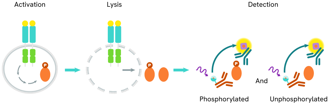
Select your location.
*e-commerce not available for this region.


This HTRF kit allows for the cell-based quantitative detection of Total HPK1
| Feature | Specification |
|---|---|
| Application | Cell Signaling |
| Sample Volume | 16 µL |
This HTRF kit allows for the cell-based quantitative detection of Total HPK1
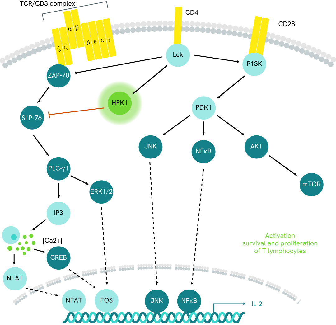
Hematopoietic progenitor kinase 1 (HPK1) is a serine/threonine kinase primarily involved in the regulation of immune cell signaling. It plays a crucial role in T-cell receptor (TCR) signaling by modulating downstream pathways that influence T-cell activation, proliferation, and differentiation. HPK1 negatively regulates TCR signaling by phosphorylating adaptor proteins such as SLP-76, which dampens T-cell responses. Additionally, HPK1 is involved in B-cell receptor (BCR) signaling, impacting B-cell activation and function. It also contributes to the innate immune response by modulating pathways in myeloid cells, such as dendritic cells and macrophages, affecting cytokine production and cell migration. Beyond immune cells, HPK1 has been implicated in the regulation of apoptosis and cell cycle progression, influencing hematopoietic cell homeostasis. Due to its regulatory roles, HPK1 is a potential therapeutic target for immune-related disorders and cancers.
| Application |
Cell Signaling
|
|---|---|
| Brand |
HTRF
|
| Buffer/Solvent |
Lysis Buffer 4
|
| Detection Modality |
HTRF
|
| Host Species |
Human
|
| Molecular Modification |
Total
|
| Product Group |
Kit
|
| Sample Volume |
16 µL
|
| Shipping Conditions |
Shipped in Dry Ice
|
| Target |
HPK1
|
| Target Class |
Phosphoproteins
|
| Technology |
TR-FRET
|
| Therapeutic Area |
Inflammation
Oncology
|
| Unit Size |
500 assay points
|
The Total HPK1 assay quantifies the expression level of HPK1 in a cell lysate. Unlike Western Blot, the assay is entirely plate-based and does not require gels, electrophoresis, or transfer. The Total HPK1 assay uses two labeled antibodies, one coupled to a donor fluorophore and the other to an acceptor. Both antibodies are highly specific for a distinct epitope on the protein. In the presence of HPK1 in a cell extract, the addition of these conjugates brings the donor fluorophore into close proximity with the acceptor and thereby generates a FRET signal. Its intensity is directly proportional to the concentration of the protein present in the sample and provides a means of assessing the protein's expression under a no-wash assay format.
The two-plate protocol involves culturing cells in a 96-well plate before lysis, then transferring lysates into a 384-well low volume detection plate before the addition of HTRF Total HPK1 detection reagents. This protocol allows for the cells' viability and confluence to be monitored.

Detection of Total HPK1 with HTRF reagents can be performed in a single plate used for culturing, stimulation, and lysis. No washing steps are required. This HTS designed protocol facilitates miniaturization while maintaining robust HTRF quality.

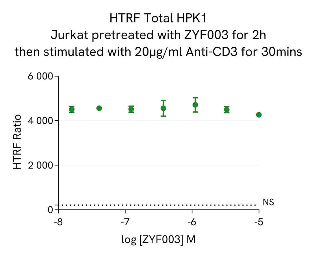
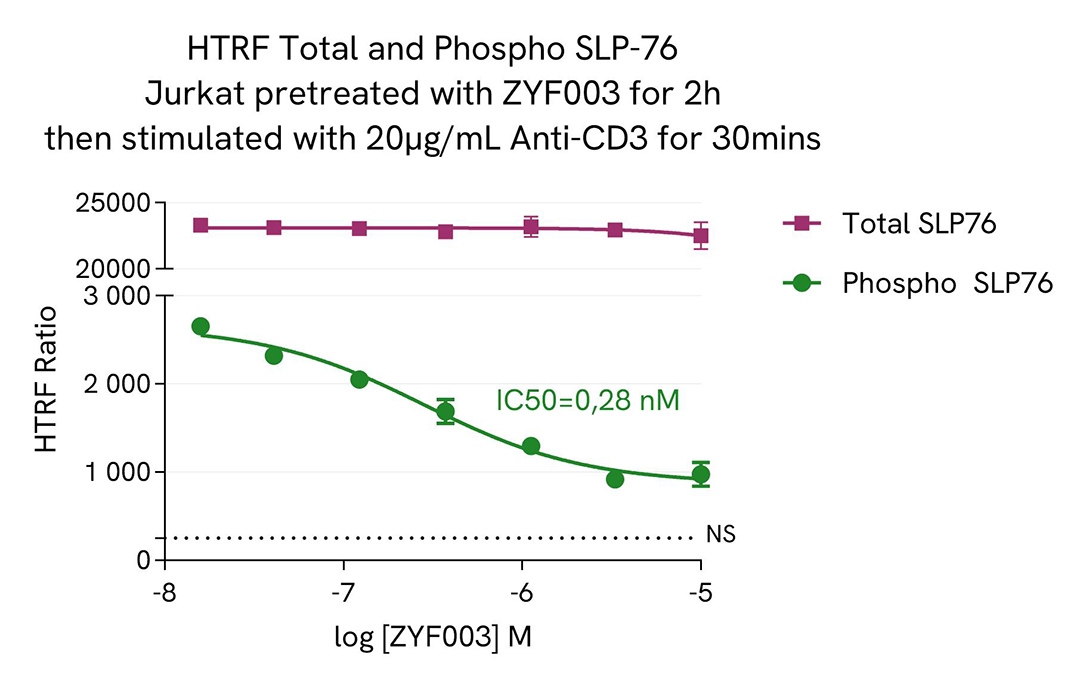
Jurkat cells (200,000 cells/well) were seeded into a 96-well culture plate and treated for 2 hours with increasing concentrations of the HPK1 inhibitor ZYF003, known to reduce the phosphorylation of downstream SLP-76. Subsequently, the cells were exposed to 20 µg/mL of anti-CD3 for 30 minutes. Following treatment, the cells were lysed with 10 µL of supplemented lysis buffer #1 (4X) for 30 minutes at room temperature with gentle shaking.
Next, 16 µL of the lysate were transferred to a 384-well low-volume white microplate, to which 4 µL of HTRF total HPK1 detection antibodies were added. Additionally, 16 µL of the lysate were analyzed using Phospho-SLP-76 (Ser376) (63ADK076PET/G/H) and Total SLP-76 (63ADK077PET/G/H) assays. Another 4 µL of lysate, supplemented with 12 µL of diluent #8, were transferred to the microplate to assess alpha-tubulin levels using the alpha-tubulin housekeeping Cellular Kit (64ATUBPET/G/H). HTRF signals were measured after overnight incubation.
The results indicated stable expression levels of Total HPK1 and Total SLP-76, alongside a decrease in Phospho-SLP-76 (Ser376) levels following ZYF003 treatment, consistent with the findings reported by Jingwen Si et al. (Cancer Cell, 2020).As expected, PMA triggered a dose-dependent increase of Phospho-cRAF (Ser43). In addition, the compound does not affect the cell viability as measured by cell viability indicator ATPLite.
Jurkat cells (200,000 cells/well) were dispensed into a 96-well plate and transfected with 5 µM siRNAs specific to HPK1, GLK, and HGK, as well as a negative control siRNA. After 48 hours, the medium was replaced with fresh culture medium, and the cells were incubated for an additional 48 hours.
Post-treatment, the cells were lysed with 10 µL of supplemented lysis buffer #1 (4X) for 30 minutes at room temperature under gentle shaking. Subsequently, 16 µL of the lysates were transferred to a 384-well low-volume white microplate, and 4 µL of HTRF Total HPK1 detection antibodies were added (following the suspension cell protocol). HTRF signals were recorded after overnight incubation.
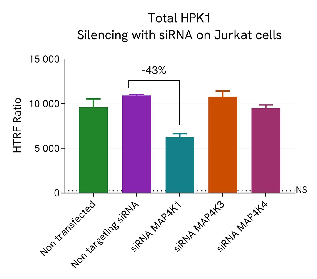
Transfection with HPK1-specific siRNA resulted in a 43% decrease in signal compared to cells transfected with the negative control siRNA, while no effect was observed with GLK and HGK siRNAs. These results demonstrate the specificity of the HTRF Total HPK1 assay, as it does not cross-react with other HPK1 family members, which share 66% and 67% amino acid identity with HPK1, respectively.
Revvity's catalog siRNA references:
The suspension cell lines Raji (lymphoblast-like cells), THP-1 (monocytes), Jurkat (T lymphocytes), and MOLT-4 (T lymphoblasts) were dispensed into a 96-well plate and lysed with 10 µL of supplemented lysis buffer #1 (4X) for 30 minutes at room temperature under gentle shaking, following the suspension cell protocol.
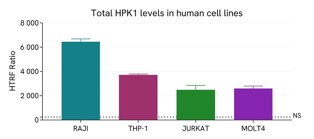
The HPK1 expression level was measured using the HTRF Total HPK1 kit. Briefly, 16 µL of cell lysate were transferred into a low-volume white microplate, followed by 4 µL of premixed HTRF detection reagents. The HTRF signal was recorded after an overnight incubation at room temperature. The dotted line indicates the non-specific HTRF signal. Cell density was optimized beforehand to ensure HTRF detection within the dynamic range of the kit (data shown for 100,000 cells/well).
The HTRF Total HPK1 assay effectively detected endogenous HPK1 in various cellular models, each expressing different levels of the protein.
Jurkat cells were cultured in a T175 flask in complete medium at 37°C, 5% CO2. After medium removal, the cells were lysed with 3 mL of supplemented lysis buffer #1 (1X) for 30 min at RT under gentle shaking.
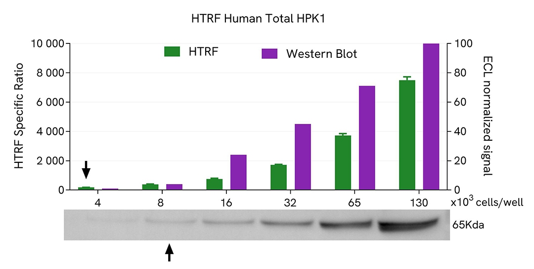
Serial dilutions of the cell lysate were performed using supplemented lysis buffer #1 (1X), and 16µL of each dilution were transferred into a 384-well small volume microplate before the addition of 4µL of HTRF Total HPK1 detection antibodies. HTRF signals were recorded after an overnight incubation.
Equal amounts of lysates were loaded into a gel for a side-by-side comparison between HTRF and Western Blot.
In these conditions, the HTRF Total HPK1 assay is 2-fold more sensitive than the Western Blot technique.
Hematopoietic progenitor kinase 1 (HPK1) plays a crucial role in T-cell receptor (TCR) signaling by acting as a negative regulator. Upon TCR engagement, HPK1 is recruited to the TCR complex where it phosphorylates adaptor proteins like SLP-76. This phosphorylation alters the function of SLP-76, reducing its ability to facilitate the assembly of signal transduction complexes at the plasma membrane. Consequently, downstream signaling events involved in T-cell activation, such as NF-κB, MAPK, and AP-1 pathways, are attenuated. HPK1 activity thus helps prevent overactivation of T-cells, maintaining immune homeostasis and preventing autoimmunity.
Are you looking for resources, click on the resource type to explore further.
Discover the versatility and precision of Homogeneous Time-Resolved Fluorescence (HTRF) technology. Our HTRF portfolio offers a...
Discover the next generation of immunoassay kits with Revvity's newly released AlphaLISA and HTRF assay targets. Our R&D...
Loading...


We are here to answer your questions.

