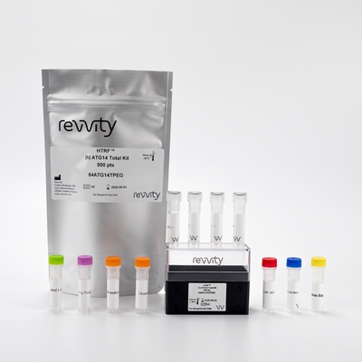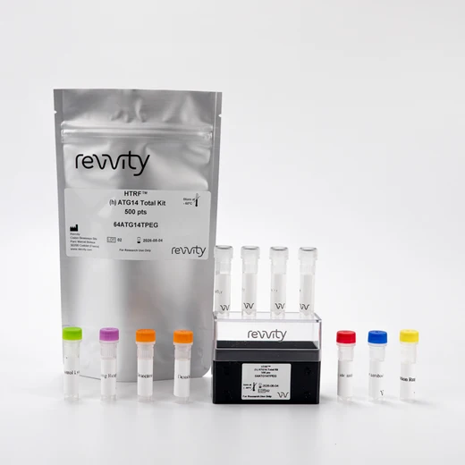

HTRF Human Total ATG14 Detection Kit, 500 Assay Points


 View All
HTRF Human Total ATG14 Detection Kit, 500 Assay Points


This HTRF kit enables the cell-based quantitative detection of ATG14 as a readout of the autophagy pathway, and can be combined with our Phospho-ATG14 Ser 29 kit.
For research use only. Not for use in diagnostic procedures. All products to be used in accordance with applicable laws and regulations including without limitation, consumption and disposal requirements under European REACH regulations (EC 1907/2006).
| Feature | Specification |
|---|---|
| Application | Cell Signaling |
| Sample Volume | 16 µL |
This HTRF kit enables the cell-based quantitative detection of ATG14 as a readout of the autophagy pathway, and can be combined with our Phospho-ATG14 Ser 29 kit.
For research use only. Not for use in diagnostic procedures. All products to be used in accordance with applicable laws and regulations including without limitation, consumption and disposal requirements under European REACH regulations (EC 1907/2006).


HTRF Human Total ATG14 Detection Kit, 500 Assay Points


HTRF Human Total ATG14 Detection Kit, 500 Assay Points


Product information
Overview
ATG14, Autophagy related protein 14, or BAKOR for Beclin 1-associated autophagy-related key regulator, is a key player in the autophagosome nucleation step in macroautophagy. Upon cellular stress, the nutrient/energy-sensitive sensors mTOR and AMPK lead to the activation of the ULK1 complex which allows phosphorylation of ATG14 on Serine 29, in turn enabling the downstream activation of the PIK3C3 autophagosome nucleation complex.
Specifications
| Application |
Cell Signaling
|
|---|---|
| Brand |
HTRF
|
| Detection Modality |
HTRF
|
| Lysis Buffer Compatibility |
Lysis Buffer 1
Lysis Buffer 4
|
| Molecular Modification |
Total
|
| Product Group |
Kit
|
| Sample Volume |
16 µL
|
| Shipping Conditions |
Shipped in Dry Ice
|
| Target Class |
Phosphoproteins
|
| Target Species |
Human
|
| Technology |
TR-FRET
|
| Unit Size |
500 Assay Points
|
Video gallery

HTRF Human Total ATG14 Detection Kit, 500 Assay Points

HTRF Human Total ATG14 Detection Kit, 500 Assay Points

How it works
Total ATG14 assay principle
The Total-ATG14 assay quantifies the expression level of ATG14 in a cell lysate. Unlike Western Blot, the assay is entirely plate-based, and does not require gels, electrophoresis, or transfer. The Total-ATG14 assay uses two labeled antibodies, one coupled to a donor fluorophore, the other to an acceptor. Both antibodies are highly specific for a distinct epitope on the protein. In presence of ATG14 in a cell extract, the addition of these conjugates brings the donor fluorophore into close proximity with the acceptor, and thereby generates a FRET signal. Its intensity is directly proportional to the concentration of the protein present in the sample, and provides a means of assessing the protein’s expression under a no-wash assay format.

Total-ATG14 two-plate assay protocol
The two-plate protocol involves culturing cells in a 96-well plate before lysis, then transferring lysates into a low volume detection plate (either HTRF 384-lv or 96-lv plate) before the addition of HTRF Total-ATG14 detection reagents. This protocol enables the cells' viability and confluence to be monitored

Total-ATG14 one-plate assay protocol
Detection of Total ATG14 with HTRF reagents can be performed in a single plate used for culturing, stimulation, and lysis. No washing steps are required. This HTS designed protocol enables miniaturization while maintaining robust HTRF quality.

Assay validation
Validation of Total and Phospho-ATG14 (Ser29) kits on the human HCT116 cell line using ULK1/2 inhibitor
Human HCT116 cells were plated in a 96-well culture-treated plate (200,000 cells/well) in complete culture medium, and incubated overnight at 37°C, 5% CO2. The cells were treated with a dose-response of MRT68921 for 4h at 37°C, 5% CO2. After culture medium removal, cells were then lysed with 25 µl of supplemented lysis buffer #4 (1X) for 30 min at RT under gentle shaking. After cell lysis, 14 µL of lysate were transferred into a 384-well low volume white microplate, and 2 µL of Activation Buffer, then 4 µL of the HTRF Total-ATG14 or Phospho-ATG14 (Ser 29) detection reagents were added. The HTRF signal was recorded after an overnight incubation at room temperature. As expected, MRT68921, a potent and dual autophagy kinase ULK1/2 inhibitor, repressed ULK1 activation, reducing autophagy initiation machinery, and leading to a dose-dependent decrease in ATG14 phosphorylation without any significant effect on the expression level of the ATG14 total protein.

Versatility of HTRF Total ATG14 assay on human cell lines
Cell lysates from various human cell lines were cultured at different densities and lysed in supplemented LB4 lysis buffer. After culture medium removal, cells were then lysed with appropriate volumes of supplemented lysis buffer #4 (1X) for 30 min at RT under gentle shaking. After cell lysis, 14 µL of lysates were transferred into a 384-well low volume white microplate, and 2 µL of Activation Buffer, then 4 µL of the HTRF Total detection reagents were added. The HTRF signal was recorded after an overnight incubation at room temperature. As expected, the Total ATG14 assay detects the human protein, as is shown by significant positive signals in several human cell lines. This demonstrates the versatility of the assay.

Simplified pathway
ATG14 signaling pathway in macro-autophagy
Cellular Autophagy is a specialized degradation and recycling process that is instrumental for cell homeostasis, being activated in response to several different stresses. There are 3 types of autophagy: macroautophagy, microautophagy, and chaperone-mediated autophagy. Macroautophagy pathways involve several key steps: initiation, nucleation, elongation of phagophores complexes, then sequestration of cytoplasmic cargos with LC3-PE recruitment followed by fusion with lysosome yielding to cargo degradations.
Biogenesis of the autophagosome is controlled by sequential and concerted actions of the so-called autophagy related proteins, ATGs, which are activated and recruited to the ER and autophagosome membranes. The recruitment of the ULK1 complex is the first event in the initiation step. ULK1 is a Ser/Thr kinase which forms a complex with the Atg13, Atg101, and FIP200 proteins. This complex is the most upstream component of the core autophagy machinery and is therefore the key initiator of autophagy in mammalian cells. ULK1 is regulated by the key nutrient/energy-sensitive kinases mTOR and AMPK, which are both able to phosphorylate ULK1 on Serine 317 and Serine 556, and directly regulate its kinase activity.
The Activated ULK1 complex then binds ATG14 via ATG13, and phosphorylates ATG14 on Serine 29. The kinase activity of phosphorylated and activated ATG14 stimulates other proteins of the PIK3C3 complex (nucleation), that is responsible for the critical step of phosphorylation of phosphatidylinositols (PI) into phosphatidylinositol-3-phosphate (PI3P). This in turn is responsible for the formation of the initial phagosomal membrane structure and later allows fixation of LC3-II using other ATG proteins (ATG16/12/5 complex), generating a support for the elongation and closing steps.
As a result, phosphorylation of the ATG14 protein on serine 29 is a key early marker of the nucleation step in the engagement of the macroautophagic process.

Resources
Are you looking for resources, click on the resource type to explore further.
Neurodegenerative diseases, such as Alzheimer’s, Parkinson’s, and Huntington’s, are complex disorders that affect millions...


How can we help you?
We are here to answer your questions.


