
HTRF Human & Mouse Total GSK3α Detection Kit, 500 Assay Points


HTRF Human & Mouse Total GSK3α Detection Kit, 500 Assay Points


The total GSK3 alpha kit monitors the cellular GSK3 alpha expression level and can be used as a normalization assay for the phospho-GSK3 alpha kit.
| Feature | Specification |
|---|---|
| Application | Cell Signaling |
| Sample Volume | 16 µL |
The total GSK3 alpha kit monitors the cellular GSK3 alpha expression level and can be used as a normalization assay for the phospho-GSK3 alpha kit.


HTRF Human & Mouse Total GSK3α Detection Kit, 500 Assay Points


HTRF Human & Mouse Total GSK3α Detection Kit, 500 Assay Points


Product information
Overview
The Total GSK3a cellular assay is used as a normalization assay with the phospho-GSK3a kit. It is optimal for metabolic research. GSK3 is involved in PI3K/AKT signaling, Wnt/ß-Catenin, Hedgehog, Notch, glycogen synthesis, and cytoskeleton polymerization, which control protein synthesis, cell proliferation, migration, inflammation, immune response, glucose regulation, and apoptosis.
Specifications
| Application |
Cell Signaling
|
|---|---|
| Brand |
HTRF
|
| Detection Modality |
HTRF
|
| Lysis Buffer Compatibility |
Lysis Buffer 1
Lysis Buffer 4
Lysis Buffer 5
|
| Molecular Modification |
Total
|
| Product Group |
Kit
|
| Sample Volume |
16 µL
|
| Shipping Conditions |
Shipped in Dry Ice
|
| Target Class |
Phosphoproteins
|
| Target Species |
Human
Mouse
|
| Technology |
TR-FRET
|
| Therapeutic Area |
Infectious Diseases
Metabolism/Diabetes
NASH/Fibrosis
|
| Unit Size |
500 assay points
|
Video gallery

HTRF Human & Mouse Total GSK3α Detection Kit, 500 Assay Points

HTRF Human & Mouse Total GSK3α Detection Kit, 500 Assay Points

How it works
Total-GSK3a assay principle
The Total-GSK3a assay quantifies the expression level of GSK3a in a cell lysate. Contrary to Western Blot, the assay is entirely plate-based and does not require gels, electrophoresis or transfer. The Total-GSK3a assay uses two labeled antibodies: one coupled to a donor fluorophore, the other to an acceptor. Both antibodies are highly specific for a distinct epitope on the protein. In presence of GSK3a in a cell extract, the addition of these conjugates brings the donor fluorophore into close proximity with the acceptor and thereby generates a FRET signal. Its intensity is directly proportional to the concentration of the protein present in the sample, and provides a means of assessing the proteins expression under a no-wash assay format.

Total-GSK3a 2-plate assay protocol
The 2 plate protocol involves culturing cells in a 96-well plate before lysis then transferring lysates to a 384-well low volume detection plate before adding Total-GSK3a HTRF detection reagents. This protocol enables the cells' viability and confluence to be monitored.

Total-GSK3a 1-plate assay protocol
Detection of total GSK3a with HTRF reagents can be performed in a single plate used for culturing, stimulation and lysis. No washing steps are required. This HTS designed protocol enables miniaturization while maintaining robust HTRF quality.

Assay validation
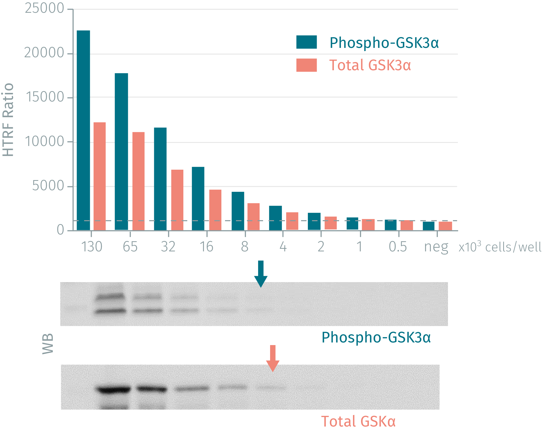
HTRF assay compared to Western Blot using phospho-GSK3a, total-GSK3a
Human MCF-7 cells were grown in a T175 flask at 37 °C, 5% CO2 until 80% confluency. After a 1.72 µM insulin stimulation for 30 min, cell culture medium was discarded and cells were lysed with 3 mL of supplemented lysis buffer for 30 min at room temperature. Soluble fractions were then collected after a 10 min centrifugation. Serial dilutions of the cell lysate were performed in the supplemented lysis buffer and 16 µL of each dilution were dispensed and analyzed side-by-side by Western Blot and by HTRF. By using HTRF phospho-GSK3a (Ser21) and total-GSK3a cellular assays, just 1,500 cells are sufficient for minimal signal detection, while 12,500 cells are needed for a Western Blot signal. The HTRF cellular assays are at least 4-fold more sensitive than the Western Blot.
Simplified pathway
GSK3 Simplified Pathway
GSK3a is active in numerous central intracellular signaling pathways and regulates important cellular processes from proliferation and immune responses, to glucose regulation and apoptosis. GSK3 kinase phosphorylation inhibits the activity of downstream targets. GSK3 is also regulated by cellular localization and by protein complex formation. In the insulin pathway, insulin activates PI3K, which phosphorylates AKT. In turn, activated AKT phosphorylates GSK3a on Ser21 and GSK3ß on Ser9, resulting in GSK-3 inactivation and inducing activation of glycogen synthase by dephosphorylation. Dephosphorylated GSK3 inactivates glycogen synthase, which converts glucose to glycogen. Thus, GSK3 is considered a versatile potential target for various disease research applications.

Resources
Are you looking for resources, click on the resource type to explore further.
Targeted protein degradation (TPD) offers a promising approach for treating neurodegenerative diseases by selectively degrading...
SDS, COAs, Manuals and more
Are you looking for technical documents related to the product? We have categorized them in dedicated sections below. Explore now.
- LanguageEnglishCountryUnited States
- LanguageFrenchCountryFrance
- LanguageGermanCountryGermany
- Resource TypeManualLanguageEnglishCountry-


Recently Viewed
How can we help you?
We are here to answer your questions.


