

HTRF BDNF Detection Kit, 500 Assay Points
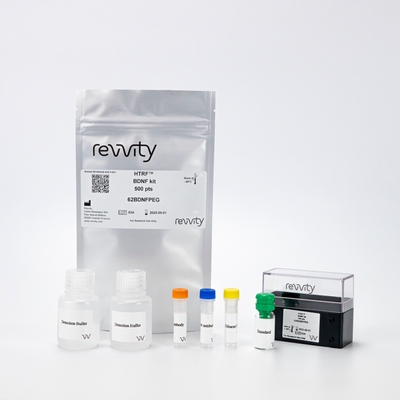

HTRF BDNF Detection Kit, 500 Assay Points
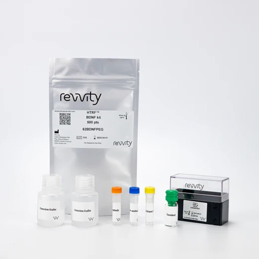


The BDNF HTRF kit is designed for the accurate quantitative measurement of Brain-Derived Neurotrophic Factor produced by cells.
| Feature | Specification |
|---|---|
| Application | Protein Quantification |
| Sample Volume | 16 µL |
The BDNF HTRF kit is designed for the accurate quantitative measurement of Brain-Derived Neurotrophic Factor produced by cells.


HTRF BDNF Detection Kit, 500 Assay Points


HTRF BDNF Detection Kit, 500 Assay Points


Product information
Overview
Brain-derived neurotrophic factor (BDNF) is a member of the neurotrophin family, which also includes nerve growth factor (NGF) and neurotrophins 3 and 4 (NT-3 and NT-4).
BDNF is one of the most widely-studied neurotrophins in healthy or diseased brains. It is secreted by neurons and glial cells, and is strongly regulated by a wide variety of endogenous and exogenous stimuli (e.g. stress, physical activity, brain injury, diet). BDNF plays a crucial role in the control of neuronal and glial development, neuroprotection, and modulation of synaptic interactions, which are critical for cognition and memory.
Decreased levels of BDNF are associated with neurodegenerative diseases such as Parkinson’s Disease, Alzheimer’s Disease, Multiple Sclerosis, and Huntington’s Disease.
Specifications
| Application |
Protein Quantification
|
|---|---|
| Brand |
HTRF
|
| Detection Modality |
HTRF
|
| Product Group |
Kit
|
| Sample Volume |
16 µL
|
| Shipping Conditions |
Shipped in Dry Ice
|
| Target Class |
Biomarkers
|
| Technology |
TR-FRET
|
| Therapeutic Area |
Neuroscience
|
| Unit Size |
500 assay points
|
Video gallery

HTRF BDNF Detection Kit, 500 Assay Points

HTRF BDNF Detection Kit, 500 Assay Points

How it works
BDNF assay principle
BDNF is measured using a sandwich immunoassay involving two specific anti-BDNF antibodies, respectively labeled with an HTRF donor and acceptor dyes. The intensity of the signal is directly proportional to the concentration of BDNF present in the sample.

BDNF assay protocol
The assay protocol, using a 384-well small volume white plate or a Revvity low volume 96-well plate (20 µL final), is described on the right. 16 µL of cell supernatant or standard are dispensed directly into the detection plate for detection by HTRF® reagents. The antibodies labeled with the HTRF donor and acceptor can be pre-mixed and added in a single dispensing step, to further streamline the assay procedure. The assay can be run in 96- to 384-well plates by simply resizing each addition volume proportionally.

Assay details
Technical specifications of BDNF kit
| Sample size | 16 µL |
|---|---|
| Final assay volume | 20 µL |
| Time to results | Overnight at RT |
| Kit components | Lyophilized standard, frozen detection antibodies, buffers & protocol |
| LOD & LOQ (in Diluent) | 5 pg/mL & 31 pg/mL |
| Range | 31 – 5,000 pg/mL |
| Calibration | NIBSC (96/534) value (IU/mL) = 0.0012 x HTRF BDNF value (pg/mL) |
| Species | Human, mouse, rat, bovine, porcine, canine |
| Specificity | No recognition of NGF, NT-3 and NT-4 |
Analytical performance
BDNF standard curve
Recombinant BDNF provided in the kit (standard) was serially diluted in diluent #5, following the procedure given in the kit’s package insert. The HTRF signal, expressed as a delta ratio, was plotted as a function of the BDNF concentrations tested.

Precision
Intra-assay (n=24)
| Sample | [BDNF] (pg/mL) | CV |
|---|---|---|
| 1 | 90 | 4% |
| 2 | 2,500 | 9% |
| 3 | 4,500 | 4% |
| Mean CV | 5.7% |
Each of the 3 samples was measured 24 times, and the % CV was calculated for each sample.
Intra-assay (n=5)
| Sample | [BDNF] (pg/mL) | CV |
|---|---|---|
| 1 | 78 | 9% |
| 2 | 312 | 7% |
| 3 | 1,250 | 4% |
| Mean CV | 6.7% |
Each of the 3 samples was measured in 5 different experiments, and the % CV was calculated for each sample.
Spike and recovery
| [BDNF] in the cell supernatant (pg/mL) | [Recombinant BDNF] added (pg/mL) | [Total BDNF] expected (pg/mL) | [Total BDNF] measured (pg/mL) | Recovery |
|---|---|---|---|---|
| 21 | 124 | 145 | 147 | 101.4% |
| 453 | 334 | 787 | 812 | 103.2% |
| 1335 | 408 | 1743 | 1730 | 99.3% |
Assay validation
Human microglial HMC3 cell line treated with IFN-γ
The human microglial cell line HMC3 was seeded in a T75 flask (3 million cells) in complete medium, and cultured until 80% confluence was reached. Cells were then treated or not with 10 ng/mL of human IFN-γ for 24h. Cell supernatants were collected, and secreted levels of BDNF were measured following the procedure given in the HTRF kit’s package insert.
In accordance with the literature, IFN-γ treated microglial cells displayed a decrease in BDNF release.

hiPSC-derived astrocytes Astro.4U treated with pro-inflammatory cytokines
Human iPSC-derived astrocytes (Astro.4U®, Ncardia) were plated in a 96-well poly-L-ornithine/laminin-coated plate and cultured for 7 days. Cells were then treated or not with a cocktail of human pro-inflammatory cytokines (10 ng/mL TNF-α + 100 ng/mL IL-1β + 100 ng/mL IFN-γ) for 24h. Cell supernatants were collected, and secreted levels of BDNF were measured following the procedure given in the HTRF kit’s package insert.
As expected, the concentration of BDNF released by astrocytes increased significantly when cells were exposed to the cocktail of pro-inflammatory cytokines.

Simplified pathway
Simplified pathway for BDNF
Astrocytes are very sensitive to their environment and respond quickly in response to CNS damage. Activated signaling pathways lead to pro- or anti-inflammatory phenotypes.
In response to some cytokines and growth factors, astrocytes activate the Jak/STAT3 pathway, which is thought to be responsible for the upregulation and secretion of many neurotrophic and neuroprotective factors promoting neuron survival and synapse repair, including BDNF.

Resources
Are you looking for resources, click on the resource type to explore further.
Dive deeper into astrocyte cell research
The release of pro-inflammatory factors by activated astrocytes has been shown to be...
SDS, COAs, Manuals and more
Are you looking for technical documents related to the product? We have categorized them in dedicated sections below. Explore now.
- LanguageEnglishCountryUnited States
- LanguageFrenchCountryFrance
- LanguageGermanCountryGermany
- Lot Number02CLot DateAugust 17, 2026
- Lot Number02ALot DateSeptember 1, 2024
- Lot Number01ELot DateSeptember 1, 2024
- Resource TypeManualLanguageEnglishCountry-


Recently viewed
How can we help you?
We are here to answer your questions.


