

HTRF Human and Mouse Phospho-ATF2 (Thr71) Detection Kit, 500 Assay Points
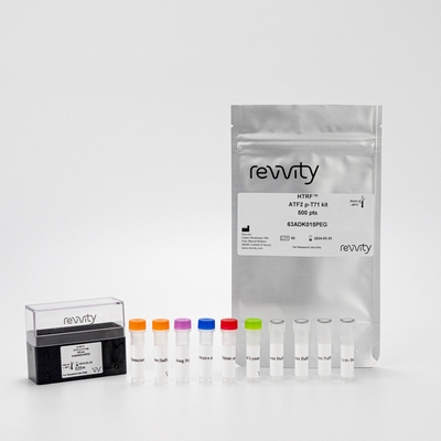

HTRF Human and Mouse Phospho-ATF2 (Thr71) Detection Kit, 500 Assay Points
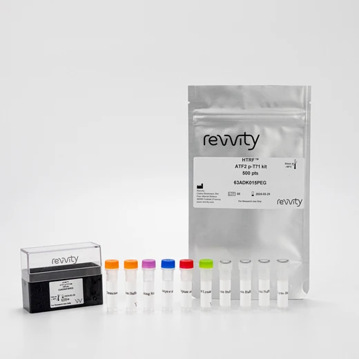


This HTRF kit enables both SAPK/JNK and p38 MAPK pathway readout, measuring ATF2 modulation phosphorylated at Threonine-71.
| Feature | Specification |
|---|---|
| Application | Cell Signaling |
| Sample Volume | 16 µL |
This HTRF kit enables both SAPK/JNK and p38 MAPK pathway readout, measuring ATF2 modulation phosphorylated at Threonine-71.


HTRF Human and Mouse Phospho-ATF2 (Thr71) Detection Kit, 500 Assay Points


HTRF Human and Mouse Phospho-ATF2 (Thr71) Detection Kit, 500 Assay Points


Product information
Overview
ATF2 (Activating transcription factor 2) is a transcription factor activated by phosphorylation on threonin residues by JNK/SAPK. The phospho-ATF2 (Thr71) kit enables the quantitative detection of transcription factor ATF2 modulation phosphorylated at Threonine-71. ATF2 is also involved in PI3K/AKT, TNF, and estrogen signaling along with insulin secretion, thyroid hormone release, substance dependence, viral carcinogenesis and infectious diseases. The simple add-and-read protocol features no wash steps for a faster analysis in biological applications like oncology, diabetes or inflammation.
Specifications
| Application |
Cell Signaling
|
|---|---|
| Brand |
HTRF
|
| Detection Modality |
HTRF
|
| Lysis Buffer Compatibility |
Lysis Buffer 1
Lysis Buffer 2
|
| Molecular Modification |
Phosphorylation
|
| Product Group |
Kit
|
| Sample Volume |
16 µL
|
| Shipping Conditions |
Shipped in Dry Ice
|
| Target Class |
Phosphoproteins
|
| Target Species |
Human
Mouse
|
| Technology |
TR-FRET
|
| Therapeutic Area |
Infectious Diseases
Metabolism/Diabetes
|
| Unit Size |
500 assay points
|
Video gallery

HTRF Human and Mouse Phospho-ATF2 (Thr71) Detection Kit, 500 Assay Points

HTRF Human and Mouse Phospho-ATF2 (Thr71) Detection Kit, 500 Assay Points

How it works
Phospho-ATF2 (Thr71) kit assay principle
The Phospho-ATF2 (Thr71) assay measures ATF2 when phosphorylated at Thr71. Contrary to Western Blot, the assay is entirely plate-based and does not require gels, electrophoresis or transfer. The Phospho-ATF2 (Thr71) assay uses 2 labeled antibodies: one with a donor fluorophore, the other one with an acceptor. The first antibody is selected for its specific binding to the phosphorylated motif on the protein, the second for its ability to recognize the protein independent of its phosphorylation state. Protein phosphorylation enables an immune-complex formation involving both labeled antibodies and which brings the donor fluorophore into close proximity to the acceptor, thereby generating a FRET signal. Its intensity is directly proportional to the concentration of phosphorylated protein present in the sample, and provides a means of assessing the protein's phosphorylation state under a no-wash assay format.

Phospho-ATF2 kit 2-plate assay protocol
The 2 plate protocol involves culturing cells in a 96-well plate before lysis then transferring lysates to a 384-well low volume detection plate before adding Phospho-ATF2 HTRF detection reagents. This protocol enables the cells' viability and confluence to be monitored.

Phospho-ATF2 kit 1-plate assay protocol
Detection of Phosphorylated ATF2 with HTRF reagents can be performed in a single plate used for culturing, stimulation and lysis. No washing steps are required. This HTS designed protocol enables miniaturization while maintaining robust HTRF quality.

Assay validation
Pharmacological validation of phospho-ATF2 (Thr71) in NIH3T3 cells
Mouse NIH3T3 cells were seeded at different cell densities in 96-well microplates, then stimulated with increasing concentrations of anisomycin for 30 minutes. Following the 2-plate assay protocol, 16 µL of lysate were transferred into a 384-well low volume white microplate before the addition of 4 µL of the HTRF phospho-ATF2 (Thr71) detection reagents. The HTRF signal was recorded after an overnight incubation.

Anisomycin dose-response and time-course in HEK293A cells
Human HEK293A suspension cells were seeded at 100,000 cells/well in a 96 well half area plate, and incubated for 24h at 37°C, 5% CO2. Then cells were stimulated with different concentrations of anisomycin for 15, 30 & 60 minutes. After lysis, 16 µL of lysate were transferred into a 384-well low volume white microplate and 4 µL of the HTRF phospho-ATF2 (Thr71) detection reagents were added. The HTRF signal was recorded after an overnight incubation.

HTRF phospho-ATF2 cellular assays compared to Western Blot
The human NIH3T3 cell line was seeded in a T175 flask and incubated a 37°C, 5% CO2 until confluency.
Serial dilutions of the cell lysate were performed in the supplemented lysis buffer, and 16 µL of each dilution were transferred into a low volume white microplate before the addition of 4 µL of HTRF phospho-ATF2 detection reagents. Equal amounts of lysates were used for a side by side comparison between HTRF and Western Blot.
Using the HTRF Phospho-ATF2 T71 assay, 3,000 cells/well were sufficient to detect a signal while 6,125 cells were needed using Western Blot with an ECL detection. These results demonstrate that the HTRF phospho-ATF2 assay is 2 times more sensitive than the Western Blot.

Simplified pathway
Function and regulation of ATF2
In response to cellular stress, such as exposure to genotoxic agents, cytokines, or UV irradiation, SAPK/JNK and p38 MAP kinases are activated and phosphorylate the transcription factor ATF-2 on residues Thr69 and Thr71. Once in the nucleus, phospho-ATF2 binds to cAMP response element (CRE) or to Activator Protein-1 (AP-1) response elements, and regulates the transcription of genes involved in cell growth, survival, and DNA damage response.

Resources
Are you looking for resources, click on the resource type to explore further.
Discover the versatility and precision of Homogeneous Time-Resolved Fluorescence (HTRF) technology. Our HTRF portfolio offers a...
This guide provides you an overview of HTRF applications in several therapeutic areas.
Loading...


Recently viewed
How can we help you?
We are here to answer your questions.


