


Total list price:
Log in to view your online price 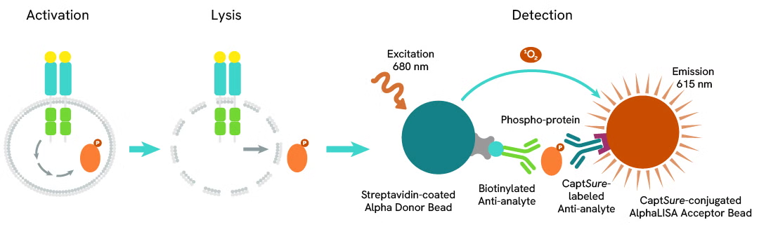
Select your location.
*e-commerce not available for this region.


The AlphaLISA™ SureFire® Ultra™ Human Phospho-Ret (Tyr905) assay is a sandwich immunoassay for quantitative detection of phospho-Ret (Tyr905) cellular lysates using Alpha Technology.
10% OFF lab essentials when you buy on Revvity.com or through your Revvity punchout with promo code.
SUPPORTU
Promo ends 5/21. Terms and conditions apply.
| Feature | Specification |
|---|---|
| Application | Cell Signaling |
| Protocol Time | 2h at RT |
| Sample Volume | 30 µL |
The AlphaLISA™ SureFire® Ultra™ Human Phospho-Ret (Tyr905) assay is a sandwich immunoassay for quantitative detection of phospho-Ret (Tyr905) cellular lysates using Alpha Technology.
Rearranged during transfection (Ret) is a receptor tyrosine kinase that governs cell growth, survival, and differentiation. Upon activation by glial cell line-derived neurotrophic factor (GDNF) family ligands, Ret triggers downstream signaling cascades, including MAPK, PI3K/AKT, and JAK/STAT. Ret fusions and activating mutations drive oncogenesis in thyroid cancer, lung adenocarcinoma, and multiple endocrine neoplasia (MEN) syndromes, making it a key target for precision therapies.
The AlphaLISA SureFire Ultra Human Phospho-Ret (Tyr905) Detection Kit is a sandwich immunoassay for the quantitative detection of phospho-Ret (Tyr905) in cellular lysates, using Alpha Technology.
Formats:
AlphaLISA SureFire Ultra kits are compatible with:
AlphaLISA SureFire Ultra kits can be used for:
| Application |
Cell Signaling
|
|---|---|
| Automation Compatible |
Yes
|
| Brand |
AlphaLISA SureFire Ultra
|
| Detection Modality |
Alpha
|
| Protocol Time |
2h at RT
|
| Sample Volume |
30 µL
|
| Shipping Conditions |
Shipped in Blue Ice
|
| Target |
Ret
|
| Target Class |
Phosphoproteins
|
| Target Species |
Human
|
| Technology |
Alpha
|
| Therapeutic Area |
Oncology
|
| Unit Size |
100 assay points
|
The Phospho-AlphaLISA SureFire Ultra assay measures a protein target when phosphorylated at a specific residue.
The assay uses two antibodies which recognize the phospho epitope and a distal epitope on the targeted protein. AlphaLISA assays require two bead types: Acceptor and Donor beads. Acceptor beads are coated with a proprietary CaptSure™ agent to specifically immobilize the assay specific antibody, labeled with a CaptSure tag. Donor beads are coated with streptavidin to capture one of the detection antibodies, which is biotinylated. In the presence of phosphorylated protein, the two antibodies bring the Donor and Acceptor beads in close proximity whereby the singlet oxygen transfers energy to excite the Acceptor bead, allowing the generation of a luminescent Alpha signal. The amount of light emission is directly proportional to the quantity of phosphoprotein present in the sample.
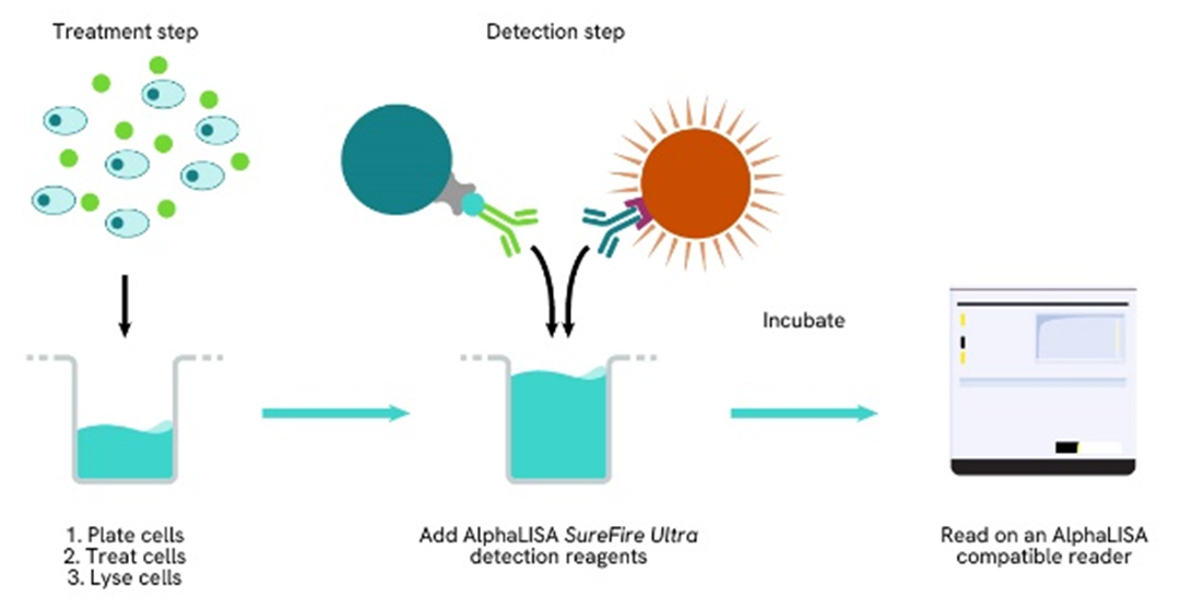
The two-plate protocol involves culturing and treating the cells in a 96-well plate before lysis, then transferring lysates into a 384-well OptiPlate™ plate before the addition of Phospho-AlphaLISA SureFire Ultra detection reagents. This protocol permits the cells viability and confluence to be monitored. In addition, lysates from a single well can be used to measure multiple targets.
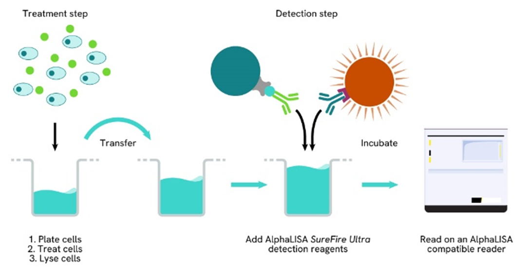
Detection of Phosphorylated target protein with AlphaLISA SureFire Ultra reagents can be performed in a single plate used for culturing, treatment, and lysis. No washing steps are required. This HTS designed protocol allows for miniaturization while maintaining AlphaLISA SureFire Ultra quality.
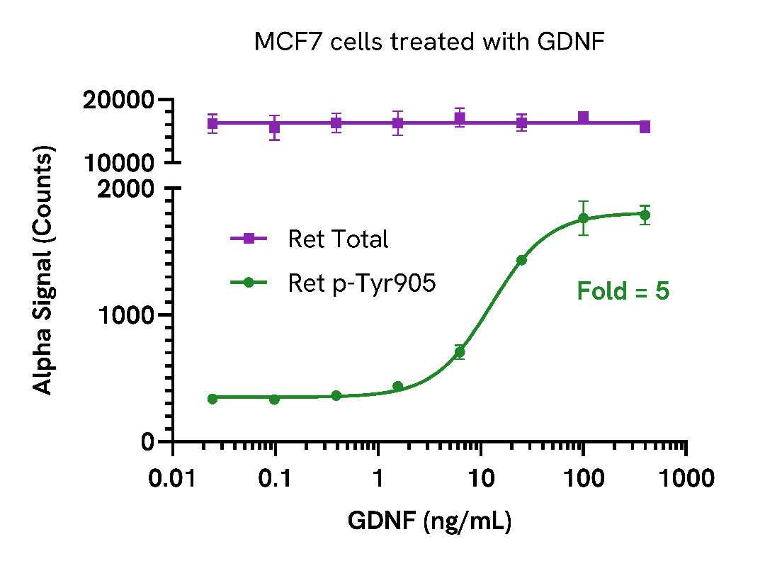
MCF7 cells were seeded in a 96-well plate (40,000 cells/well) in complete media, and incubated overnight at 37°C, 5% CO2. The cells were then starved overnight in serum-free media and treated with increasing concentrations of GDNF for 15 minutes.
After treatment, the cells were lysed with 100 µL of Lysis Buffer for 10 minutes at RT with shaking (350 rpm). Ret Phospho (Tyr905) and Total levels were evaluated using respective AlphaLISA SureFire Ultra assays. For the detection step, 10 µL of cell lysate (approximately 4,000 cells) was transferred into a 384-well white OptiPlate, followed by 5 µL of Acceptor mix and incubated for 1 hour at RT. Finally, 5 µL of Donor mix was then added to each well and incubated for 1 hour at RT in the dark. The plate was read on an Envision using standard AlphaLISA settings.
As expected, GDNF triggered a dose-dependent increase in the levels of Ret Phospho (Tyr905) while Ret Total levels remained unchanged.
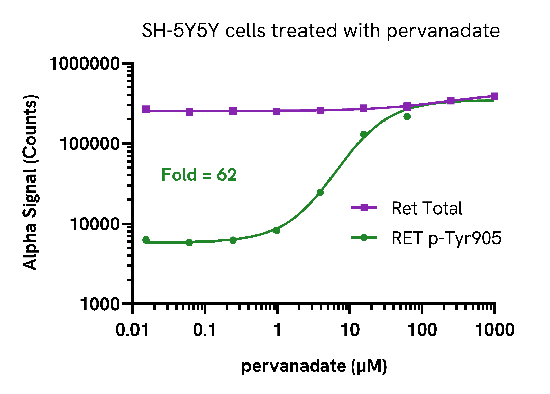
SH-SY5Y cells were seeded in a 96-well plate (40,000 cells/well) and incubated overnight at 37°C, 5% CO2. The cells were treated with 12.5 µM Retinoic Acid for 18 hours and then increasing concentrations of pervanadate for 30 minutes.
After treatment, the cells were lysed with 100 µL of Lysis Buffer for 10 minutes at RT with shaking (350 rpm). Ret Phospho (Tyr905) and Total levels were evaluated using respective AlphaLISA SureFire Ultra assays. For the detection step, 10 µL of cell lysate (approximately 4,000 cells) was transferred into a 384-well white OptiPlate, followed by 5 µL of Acceptor mix and incubated for 1 hour at RT. Finally, 5 µL of Donor mix was then added to each well and incubated for 1 hour at RT in the dark. The plate was read on an Envision using standard AlphaLISA settings.
As expected, pervanadate caused a dose-dependent increase in the levels of Ret Phospho (Tyr905) while Ret Total levels remained unchanged.
Are you looking for technical documents related to the product? We have categorized them in dedicated sections below. Explore now.


We are here to answer your questions.

