


Total list price:
Log in to view your online price 
Select your location.
*e-commerce not available for this region.


The AlphaLISA™ SureFire® Ultra™ Human and Mouse Total GSPT1 assay is a sandwich immunoassay for quantitative detection of total GSPT1 cellular lysates using Alpha Technology.
| Feature | Specification |
|---|---|
| Application | Cell Signaling |
| Protocol Time | 2h at RT |
| Sample Volume | 30 µL |
The AlphaLISA™ SureFire® Ultra™ Human and Mouse Total GSPT1 assay is a sandwich immunoassay for quantitative detection of total GSPT1 cellular lysates using Alpha Technology.
G1 to S phase transition 1 (GSPT1), also known as eRF3a (eukaryotic Release Factor 3a), is a translation termination factor that interacts with eRF1 to regulate mRNA stability and degradation. It plays a crucial role in cell cycle progression, proliferation, and protein synthesis termination. GSPT1 contains a GTPase domain essential for its activity and interacts with various factors, including PABP and components of the mTOR signaling pathway. Dysregulation of GSPT1 is implicated in hematologic malignancies and solid tumors, influencing cell proliferation, apoptosis, and metastasis. Overexpression of GSPT1 can lead to enhanced cell growth and survival, contributing to drug resistance in some cancers. Targeting GSPT1 through protein degradation strategies has emerged as a promising therapeutic approach in cancer treatment.
The AlphaLISA SureFire Ultra Human and Mouse Total GSPT1 Detection Kit is a sandwich immunoassay for the quantitative detection of total GSPT1 in cellular lysates, using Alpha Technology.
Formats:
AlphaLISA SureFire Ultra kits are compatible with:
AlphaLISA SureFire Ultra kits can be used for:
| Application |
Cell Signaling
|
|---|---|
| Automation Compatible |
Yes
|
| Brand |
AlphaLISA SureFire Ultra
|
| Detection Modality |
Alpha
|
| Protocol Time |
2h at RT
|
| Sample Volume |
30 µL
|
| Shipping Conditions |
Shipped in Blue Ice
|
| Target |
GSPT1
|
| Target Class |
Phosphoproteins
|
| Target Species |
Human
Mouse
|
| Technology |
Alpha
|
| Therapeutic Area |
Oncology
|
| Unit Size |
100 assay points
|
The Total-AlphaLISA SureFire Ultra assay measures the expression level of a protein target in a cell lysate.
The Total-AlphaLISA SureFire Ultra assay uses two antibodies which recognize two different distal epitopes on the targeted protein. AlphaLISA assays require two bead types: Acceptor and Donor beads. Acceptor beads are coated with a proprietary CaptSure™ agent to specifically immobilize the assay specific antibody, labeled with a CaptSure tag. Donor beads are coated with streptavidin to capture one of the detection antibodies, which is biotinylated. In the presence of targeted protein, the two antibodies bring the Donor and Acceptor beads in close proximity whereby the singlet oxygen transfers energy to excite the Acceptor bead, allowing the generation of a luminescent Alpha signal. The amount of light emission is directly proportional to the quantity of protein present in the sample.
The two-plate protocol involves culturing and treating the cells in a 96-well plate before lysis, then transferring lysates into a 384-well OptiPlate™ plate before the addition of Total-AlphaLISA SureFire Ultra detection reagents. This protocol permits the cells viability and confluence to be monitored. In addition, lysates from a single well can be used to measure multiple targets.

Detection of Total target protein with AlphaLISA SureFire Ultra reagents can be performed in a single plate used for culturing, treatment, and lysis. No washing steps are required. This HTS designed protocol allows for miniaturization while maintaining AlphaLISA SureFire Ultra quality.

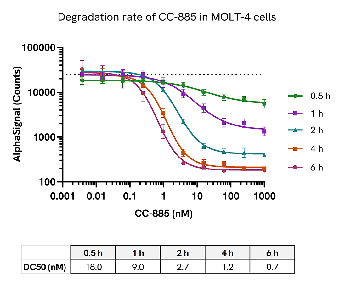
MOLT-4 cells were seeded in a 96-well plate (250,000 cells/well) in HBSS + 0.1% BSA. The cells were treated with increasing concentrations of CC-885 for up to 6 hours.
After treatment, the cells were lysed with 50 µL of 5X Lysis Buffer for 10 minutes at RT with shaking (350 rpm). GSPT1 levels were evaluated using the AlphaLISA SureFire Ultra assay. For the detection step, 10 µL of cell lysate (approximately 10,000 cells) was transferred into a 384-well white OptiPlate, followed by 5 µL of Acceptor mix and incubated for 1 hour at RT. Finally, 5 µL of Donor mix was then added to each well and incubated for 1 hour at RT in the dark. The plate was read on an Envision using standard AlphaLISA settings.
The DC50 decreases with the length of CC-885 treatment, demonstrating an increase of potency over time.
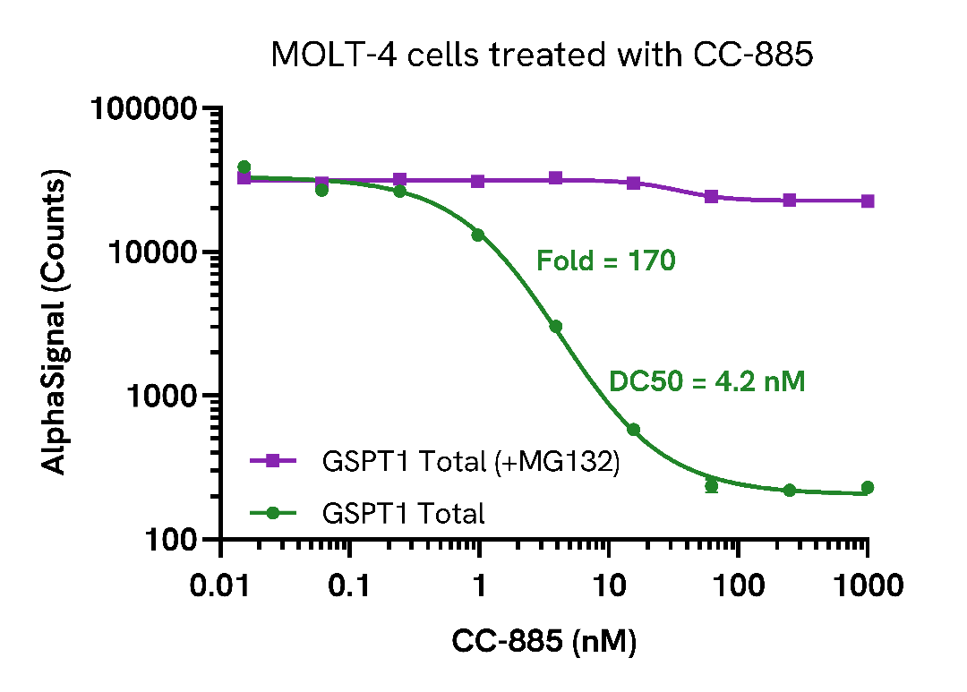
MOLT-4 cells were seeded in a 96-well plate (250,000 cells/well) in HBSS + 0.1% BSA. The cells were treated for 1 hour with 5 µM MG132, followed by increasing concentrations of CC-885 for 2 hours.
After treatment, the cells were lysed with 50 µL of 5X Lysis Buffer for 10 minutes at RT with shaking (350 rpm). GSPT1 levels were evaluated using the AlphaLISA SureFire Ultra assay. For the detection step, 10 µL of cell lysate (approximately 10,000 cells) was transferred into a 384-well white OptiPlate, followed by 5 µL of Acceptor mix and incubated for 1 hour at RT. Finally, 5 µL of Donor mix was then added to each well and incubated for 1 hour at RT in the dark. The plate was read on an Envision using standard AlphaLISA settings.
As expected, CC-885 induced a dose-dependent decrease in GSPT1 Total levels. The lack of protein degradation in the presence of MG132 confirmed that the degradation was driven by the ubiquitin-proteosome system.
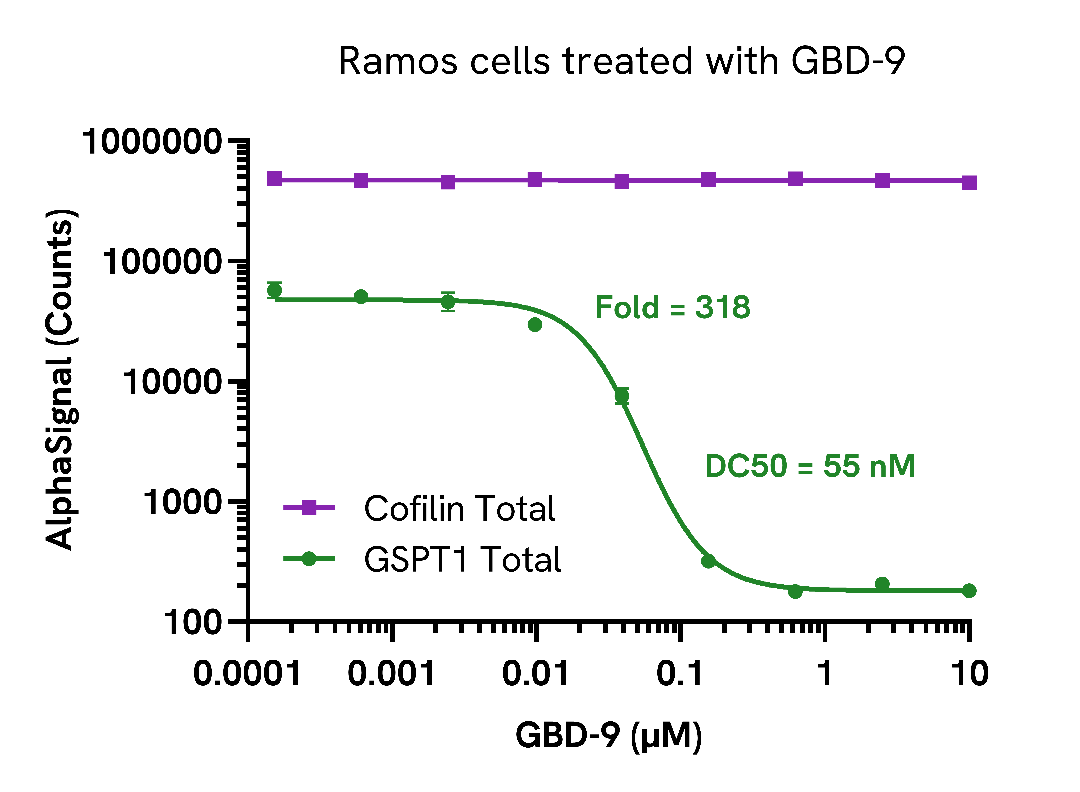
Ramos cells were seeded in a 96-well plate (100,000 cells/well) in complete medium. The cells were treated with increasing concentrations of GBD-9 for 16 hours.
After treatment, the cells were spun down, washed with HBSS and lysed with 100 µL of Lysis Buffer for 10 minutes at RT with shaking (350 rpm). GSPT1 and Cofilin Total levels were evaluated using respective AlphaLISA SureFire Ultra assays. For the detection step, 10 µL of cell lysate (approximately 10,000 cells) was transferred into a 384-well white OptiPlate, followed by 5 µL of Acceptor mix and incubated for 1 hour at RT. Finally, 5 µL of Donor mix was then added to each well and incubated for 1 hour at RT in the dark. The plate was read on an Envision using standard AlphaLISA settings.
As expected, GBD-9 treatment resulted in a dose-dependent decrease in the levels of Total GSPT1.
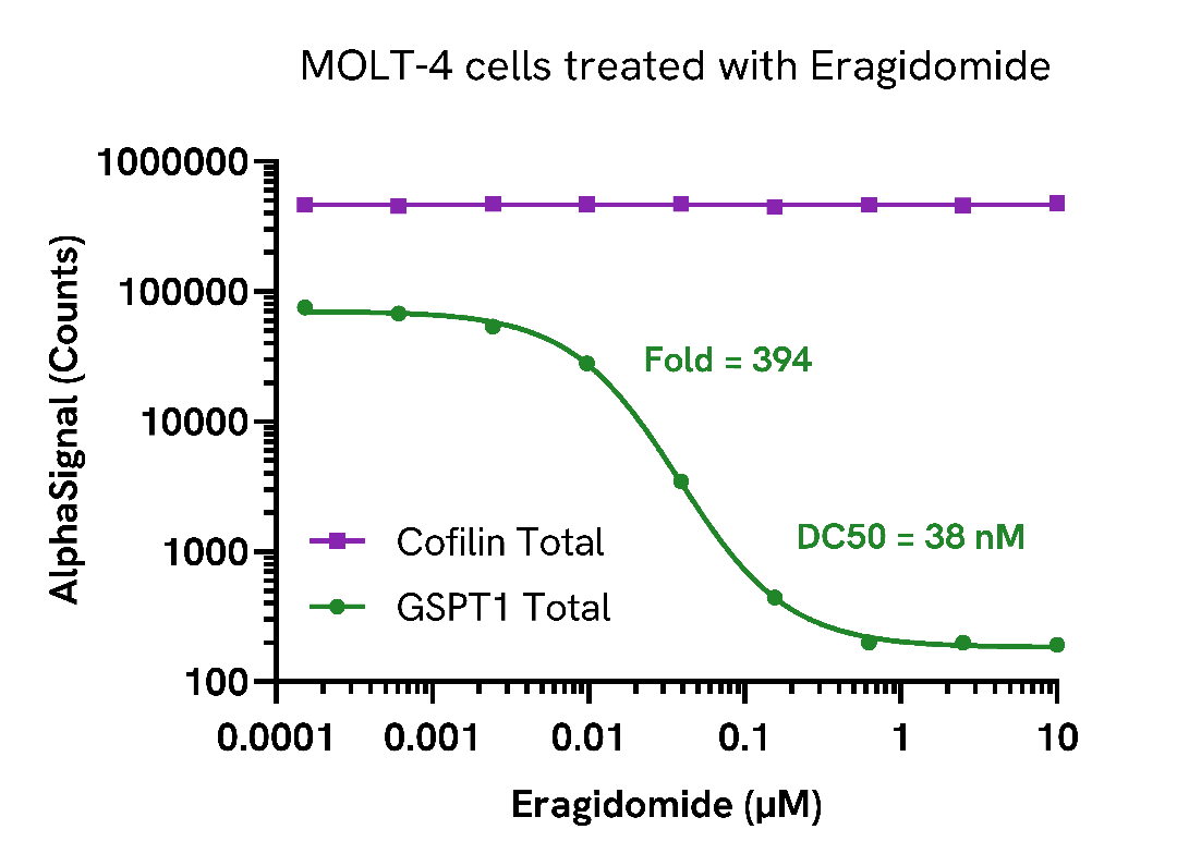
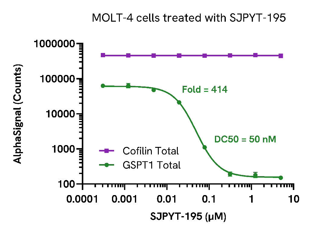
MOLT-4 cells were seeded in a 96-well plate (250,000 cells/well) in complete medium. The cells were treated with increasing concentrations of Eragidomide or SJPYT-195 for 16 hours.
After treatment, the cells were spun down, washed with HBSS and lysed with 100 µL of Lysis Buffer for 10 minutes at RT with shaking (350 rpm). GSPT1 and Cofilin Total levels were evaluated using respective AlphaLISA SureFire Ultra assays. For the detection step, 10 µL of cell lysate (approximately 25,000 cells) was transferred into a 384-well white OptiPlate, followed by 5 µL of Acceptor mix and incubated for 1 hour at RT. Finally, 5 µL of Donor mix was then added to each well and incubated for 1 hour at RT in the dark. The plate was read on an Envision using standard AlphaLISA settings.
As expected, treatment with Eragidomide or SJPYT-195 resulted in a dose-dependent decrease in the total levels of GSPT1.
Selectivity of the Total GSPT1 assay was assessed by assaying recombinant GSPT1 protein.
Dilutions of recombinant GSPT1 protein (Abcam ab12287) was prepared in Lysis Buffer and evaluated using the AlphaLISA SureFire Ultra assay. For the detection step, 10 µL of protein was transferred into a 384-well white OptiPlate, followed by 5 µL of Acceptor mix and incubated for 1 hour at room temperature. Finally, 5 µL of Donor mix was then added to each well and incubated for 1 hour at RT in the dark. The plate was read on an Envision using standard AlphaLISA settings.

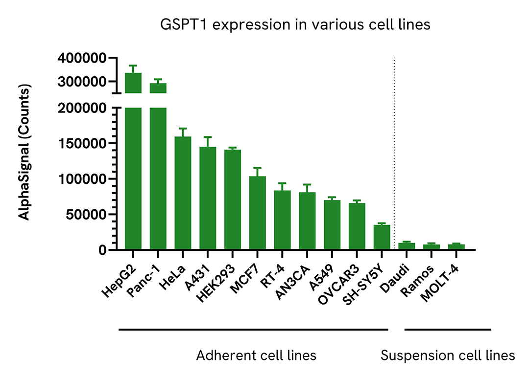
Adherent cells were seeded at 40,000 cells/well in a 96-well culture plate in complete medium and incubated overnight at 37°C, 5% CO2. Cells were lysed with 100 µL of Lysis Buffer for 10 minutes at RT with shaking (350 rpm). Suspension cells were seeded at 100,000 cells/well (200 µL/well) in a 96-well culture plate in HBSS + 0.1% BSA and lysed with 50 µL of 5X Lysis Buffer for 10 minutes at RT with shaking (350 rpm).
Total GSPT1 levels were evaluated by AlphaLISA SureFire Ultra. For the detection step, 10 µL of cell lysate (4,000 cells) was transferred into a 384-well white OptiPlate, followed by 5 µL of Acceptor Mix and incubated for 1 hour at RT. Finally, 5 µL of Donor Mix was then added to each well and incubated for 1 hour at RT in the dark. The plate was read on an Envision using standard AlphaLISA settings.
GSPT1 is highly expressed in most cell lines tested.
Loading...


We are here to answer your questions.

