


Total list price:
Log in to view your online price 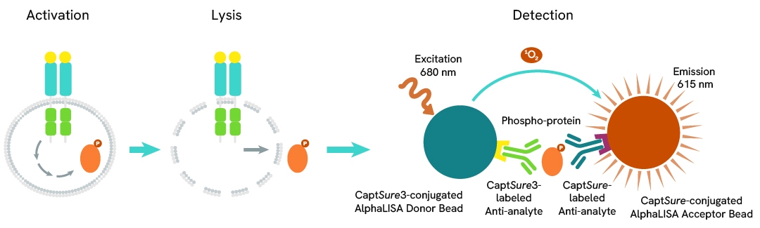
Select your location.
*e-commerce not available for this region.


The AlphaLISA™ SureFire® Human and Mouse Phospho-STAT5 (Tyr694/699) assay is a sandwich immunoassay for quantitative detection of phospho-STAT5 (Tyr694/699) in cellular lysates using Alpha Technology.
| Feature | Specification |
|---|---|
| Application | Cell Signaling |
| Protocol Time | 2h at RT |
The AlphaLISA™ SureFire® Human and Mouse Phospho-STAT5 (Tyr694/699) assay is a sandwich immunoassay for quantitative detection of phospho-STAT5 (Tyr694/699) in cellular lysates using Alpha Technology.
STAT5, comprising STAT5A and STAT5B, is activated by JAK kinases in response to cytokine signaling. Upon phosphorylation, STAT5 forms dimers and translocates to the nucleus, inducing transcription of genes involved in proliferation, differentiation, and survival. Overactive STAT5 drives cancers like leukemia and breast cancer, while its dysregulation contributes to autoimmune diseases by promoting excessive immune responses.
The AlphaLISA SureFire Biotin-Free Human and Mouse Phospho-STAT5 (Tyr694/699) Detection Kit is a sandwich immunoassay for the quantitative detection of phospho-STAT5 in cellular lysates, using Alpha Technology.
Formats:
AlphaLISA SureFire Biotin Free kits are compatible with:
AlphaLISA SureFire Biotin Free kits can be used for:
| Application |
Cell Signaling
|
|---|---|
| Automation Compatible |
Yes
|
| Brand |
AlphaLISA SureFire Biotin-Free
|
| Detection Modality |
Alpha
|
| Protocol Time |
2h at RT
|
| Shipping Conditions |
Shipped in Blue Ice
|
| Target |
STAT5
|
| Target Class |
Phosphoproteins
|
| Target Species |
Human
Mouse
|
| Technology |
Alpha
|
| Therapeutic Area |
Autoimmunity
Oncology
|
| Unit Size |
500 assay points
|
The Phospho-AlphaLISA SureFire Biotin Free assay measures a protein target when phosphorylated at a specific residue.
The assay uses two antibodies which recognize the phospho epitope and a distal epitope on the targeted protein. AlphaLISA assays require two bead types: Acceptor and Donor beads. Acceptor beads are coated with a proprietary CaptSure™ agent to specifically immobilize the assay specific antibody, labeled with a CaptSure tag. Donor beads are coated with a proprietary CaptSure 3 agent to capture one of the detection antibodies, which is labeled with CaptSure 3 tag. In the presence of phosphorylated protein, the two antibodies bring the Donor and Acceptor beads in close proximity whereby the singlet oxygen transfers energy to excite the Acceptor bead, allowing the generation of a luminescent Alpha signal. The amount of light emission is directly proportional to the quantity of phosphoprotein present in the sample.
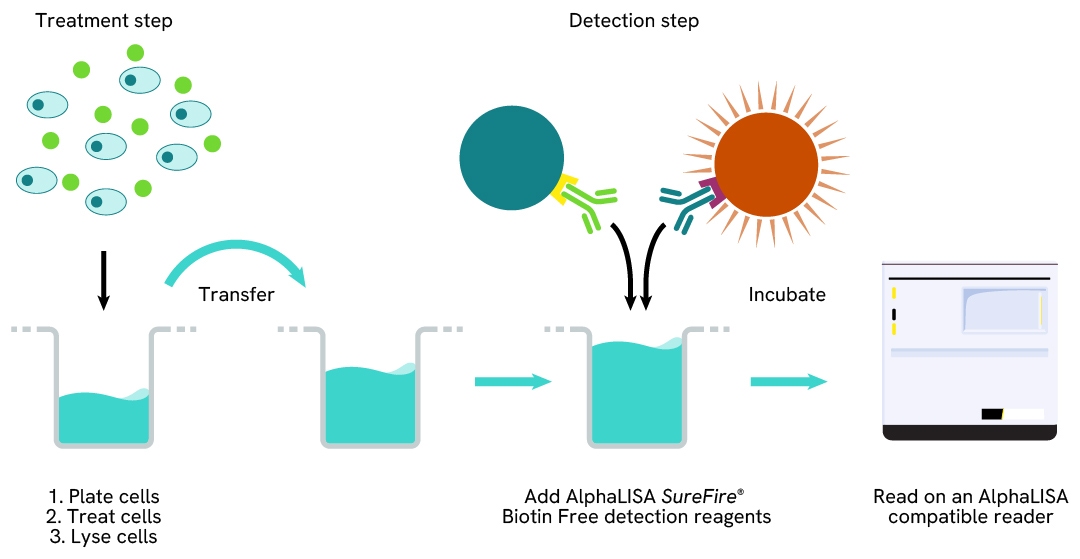
The two-plate protocol involves culturing and treating the cells in a 96-well plate before lysis, then transferring lysates into a 384-well Optiplate™ plate before the addition of Phospho-AlphaLISA SureFire Biotin Free detection reagents. This protocol permits the cells viability and confluence to be monitored. In addition, lysates from a single well can be used to measure multiple targets.
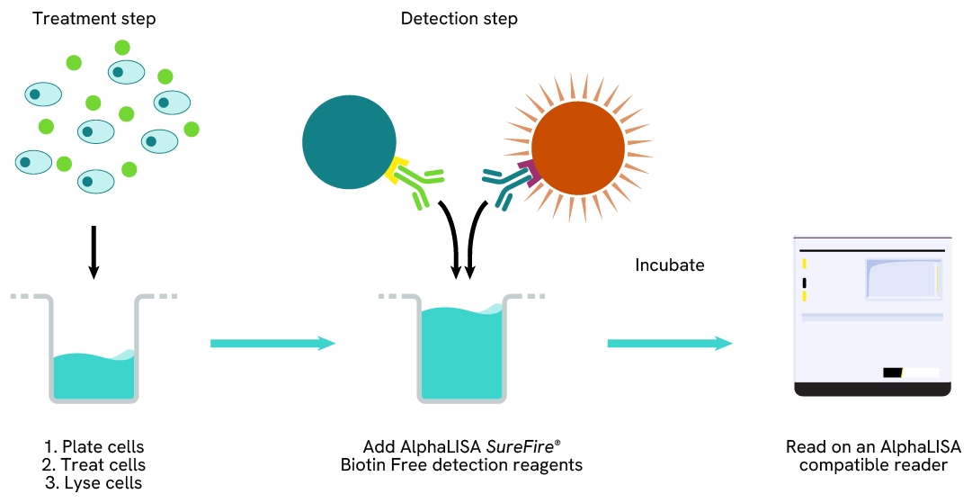
Detection of Phosphorylated target protein with AlphaLISA SureFire Biotin Free reagents can be performed in a single plate used for culturing, treatment, and lysis. No washing steps are required. This HTS designed protocol allows for miniaturization while maintaining robust AlphaLISA SureFire quality.
THP-1 cells were seeded in a 96-well plate (100,000 cells/well) in complete RPMI 1640 medium containing 100 nM of PMA for 24 hours at 37°C, 5% CO2. The THP-1 derived macrophages were starved for 2 hours and then treated with increasing concentrations of IFNα or IFNγ for 20 minutes.
After treatment, the cells were lysed with the addition of 50 µL of 5X Lysis Buffer for 10 minutes at RT with shaking (350 rpm). STAT5 Phospho (Tyr694/699) and Total levels were evaluated using respective AlphaLISA SureFire Biotin Free assays. For the detection step, the lysate was further diluted 1:2 with Lysis Buffer, 10 µL of cell lysate (approximately 2,000 cells) were transferred into a 384-well white OptiPlate, followed by 5 µL of Acceptor mix and incubated for 1 hour at RT. Finally, 5 µL of Donor mix was then added to each well and incubated for 1 hour at RT in the dark. The plate was read on an Envision using standard AlphaLISA settings.
As expected, IFNα and IFNγ triggered a dose-dependent increase in the levels of Phospho STAT5 (Tyr694/699) while Total STAT5 levels remained unchanged.


Are you looking for resources, click on the resource type to explore further.
The definitive guide for setting up a successful AlphaLISA SureFire Ultra assay
Several biological processes are regulated by...
Discover Alpha SureFire® Ultra™ assays, the no-wash cellular kinase assays leveraging Revvity's exclusive bead-based technology...
Featuring homogeneous detection of cell-based proteins, this note shows you how to utilize an innovative system optimized for...
This document includes detailed tables listing HTRF™, AlphaLISA™ SureFire® Ultra™, and Alpha SureFire® Ultra™ Multiplex assays...
Loading...


We are here to answer your questions.

