


Total list price:
Log in to view your online price USD 48,363.00
Select your location.
*e-commerce not available for this region.


| Feature | Specification |
|---|---|
| Application | Cell Signaling |
| Protocol Time | 2h at RT |
| Sample Volume | 10 µL |
Interferon regulatory factor 5 (IRF5) is a transcription factor involved in innate immunity and inflammatory responses, regulating the expression of type I interferons (IFN-alpha and IFN-beta) and pro-inflammatory cytokines. Activated by pattern recognition receptors such as Toll-like receptors (TLR), MDA5, or STING in response to viral infection, IRF5 plays a crucial role in antiviral defense. In its inactive form, IRF5 resides in the cytoplasm of uninfected cells. Upon activation, it undergoes phosphorylation, dimerization, and nuclear localization to drive gene expression. Dysregulation of IRF5 is linked to autoimmune diseases such as lupus and rheumatoid arthritis, as well as inflammatory cancers, where it modulates tumor-associated macrophages.
The AlphaLISA SureFire Biotin-Free Human and Mouse Total IRF5 Detection Kit is a sandwich immunoassay for the quantitative detection of total IRF5 in cellular lysates, using Alpha Technology.
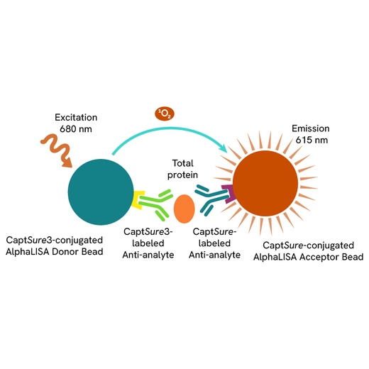
The Total-AlphaLISA Surefire Ultra Biotin Free assay measures the expression level of a protein target in a cell lysate.
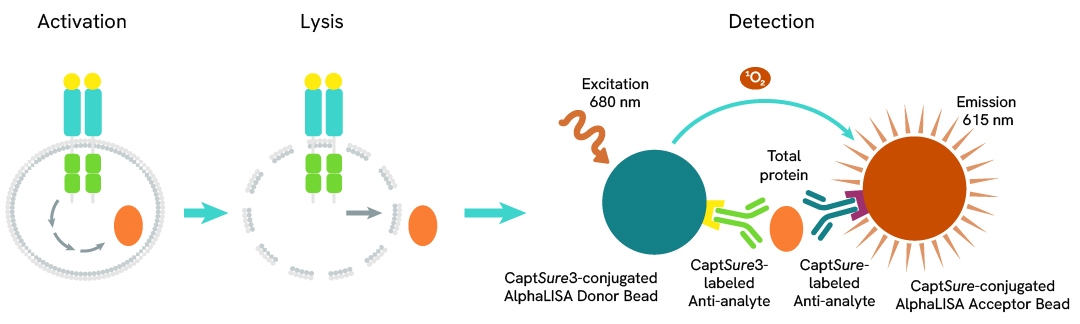
The Total-AlphaLISA Surefire Ultra assay uses two antibodies which recognize two different distal epitopes on the targeted protein. AlphaLISA assays require two bead types: Acceptor and Donor beads. Acceptor beads are coated with a proprietary CaptSure™ agent to specifically immobilize the assay specific antibody, labeled with a CaptSure tag. Donor beads are coated with a proprietary CaptSure 3 agent to capture one of the detection antibodies, which is labeled with CaptSure 3 tag. In the presence of targeted protein, the two antibodies bring the Donor and Acceptor beads in close proximity whereby the singlet oxygen transfers energy to excite the Acceptor bead, allowing the generation of a luminescent Alpha signal. The amount of light emission is directly proportional to the quantity of protein present in the sample.
The two-plate protocol involves culturing and treating the cells in a 96-well plate before lysis, then transferring lysates into a 384-well Optiplate™ plate before the addition of Total-AlphaLISA SureFire Ultra Biotin Free detection reagents. This protocol permits the cells viability and confluence to be monitored. In addition, lysates from a single well can be used to measure multiple targets.
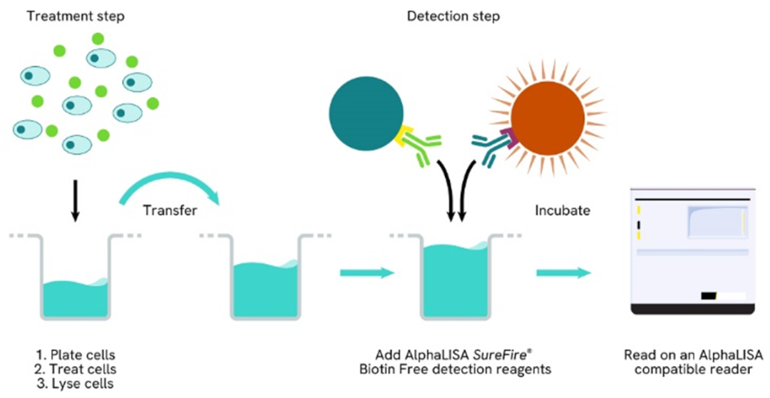
Detection of Total target protein with AlphaLISA SureFire Ultra Biotin Free reagents can be performed in a single plate used for culturing, treatment, and lysis. No washing steps are required. This HTS designed protocol allows for miniaturization while maintaining robust AlphaLISA SureFire Ultra quality.
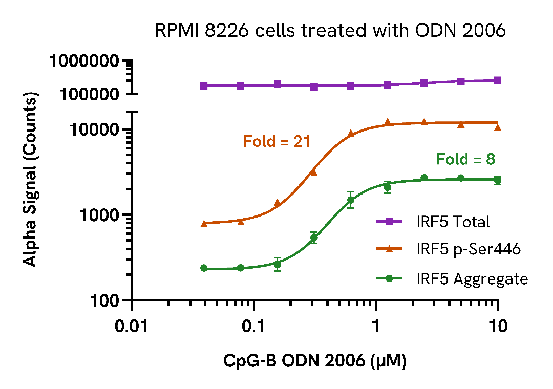
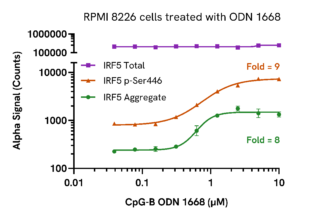
RPMI 8226 cells were seeded in a 96-well plate (200,000 cells/well) in complete medium and incubated for approximately 10 minutes at 37°C, 5% CO2. The cells were treated with increasing concentrations of CpG-B ODN 2006 or ODN 1668 (MedChem Express, HY-150218, HY-150726 respectively) for 3 hours.
After treatment, the cells were spun down at 1200 rpm for 5 minutes, supernatant was removed and cells were lysed with 50 µL of Lysis Buffer for 10 minutes at RT with shaking (350 rpm). Aggregate, Phospho (Ser446) and Total IRF5 levels were evaluated using respective AlphaLISA SureFire Biotin Free assays. For the detection step, 10 µL of cell lysate (approximately 40,000 cells) were transferred into a 384-well white OptiPlate, followed by 5 µL of Acceptor mix and incubated for 1 hour at RT. Finally, 5 µL of Donor mix was then added to each well and incubated for 1 hour at RT in the dark. The plate was read on an Envision using standard AlphaLISA settings.
As expected, CpG-B triggered a dose-dependent increase in Aggregate and Phospho (Ser446) IRF5 while Total levels remained unchanged.
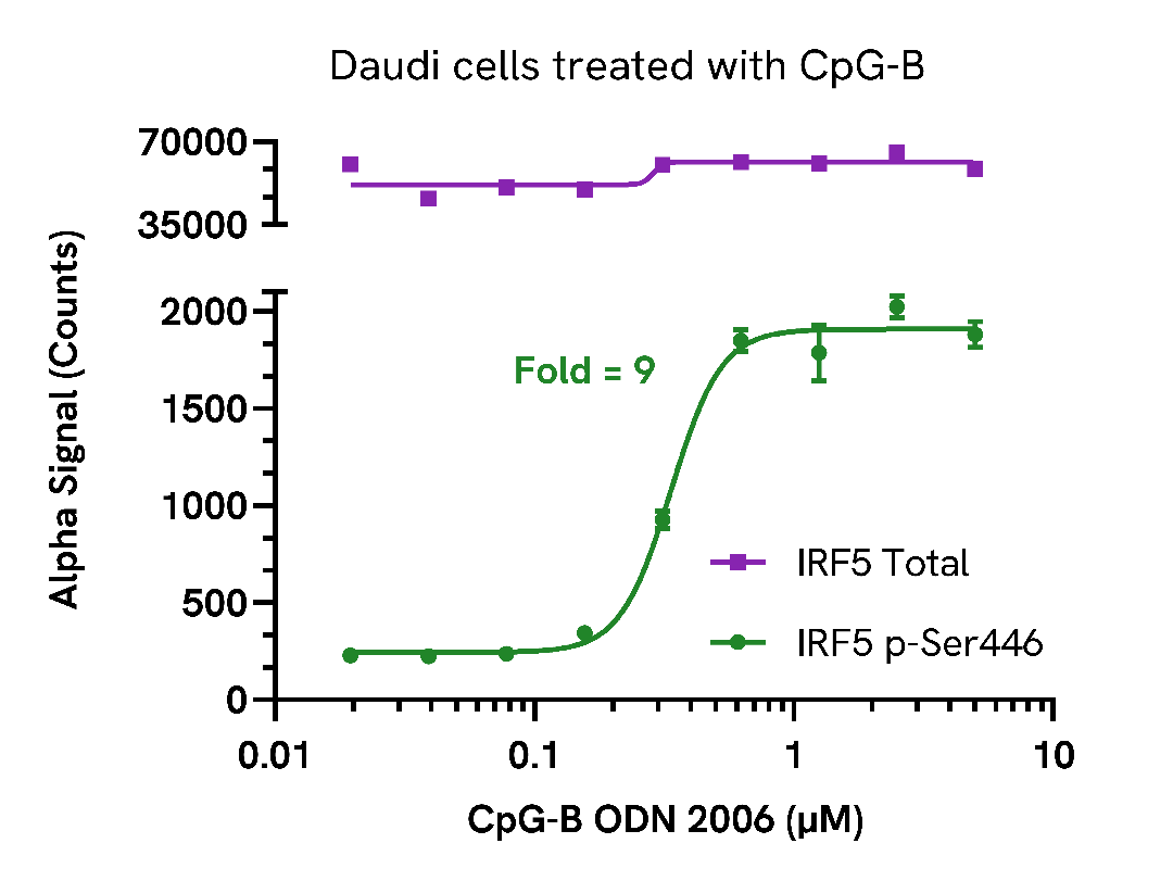
Daudi cells were seeded in a 96-well plate (400,000 cells/well) in complete medium and incubated for approximately 10 minutes at 37°C, 5% CO2. The cells were treated with increasing concentrations of CpG-B ODN 2006 (MedChem Express, HY-150218) for 3 hours.
After treatment, the cells were spun down at 1200 rpm for 5 minutes, supernatant was removed and cells were lysed with 50 µL of Lysis Buffer for 10 minutes at RT with shaking (350 rpm). Phospho (Ser446) and Total IRF5 levels were evaluated using respective AlphaLISA SureFire Biotin Free assays. For the detection step, 10 µL of cell lysate (approximately 80,000 cells) was transferred into a 384-well white OptiPlate, followed by 5 µL of Acceptor mix and incubated for 1 hour at RT. Finally, 5 µL of Donor mix was then added to each well and incubated for 1 hour at RT in the dark. The plate was read on an Envision using standard AlphaLISA settings.
As expected, CpG-B triggered a dose-dependent increase in Phospho (Ser446) IRF5 while Total levels remained unchanged.
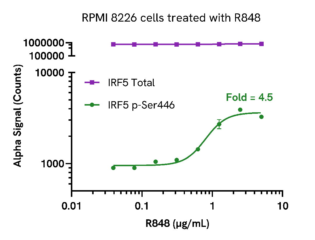
RPMI 8226 cells were seeded in a 96-well plate (400,000 cells/well) in complete medium and incubated for approximately 10 minutes at 37°C, 5% CO2. The cells were treated with increasing concentrations of R848 (InvivoGen) for 4 hours.
After treatment, the cells were spun down at 1200 rpm for 5 minutes, supernatant was removed and cells were lysed with 50 µL of Lysis Buffer for 10 minutes at RT with shaking (350 rpm). Phospho (Ser446) and Total IRF5 levels were evaluated using respective AlphaLISA SureFire Biotin Free assays. For the detection step, 10 µL of cell lysate (approximately 80,000 cells) was transferred into a 384-well white OptiPlate, followed by 5 µL of Acceptor mix and incubated for 1 hour at RT. Finally, 5 µL of Donor mix was then added to each well and incubated for 1 hour at RT in the dark. The plate was read on an Envision using standard AlphaLISA settings.
Treatment with a TLR7/8 agonist, R848, induced a dose-dependent increase in the levels of Phospho IRF5 while Total levels remained unchanged.
THP-1 cells were seeded in a 96-well plate (200,000 cells/well) in complete medium and incubated for approximately 10 minutes at 37°C, 5% CO2. The cells were treated with increasing concentrations of Calyculin A for 3 hours.
After treatment, the cells were spun down at 1200 rpm for 5 minutes, supernatant was removed, and cells were lysed with 50 µL of Lysis Buffer for 10 minutes at RT with shaking (350 rpm). Aggregate, Phospho (Ser446) and Total IRF5 levels were evaluated using respective AlphaLISA SureFire Biotin Free assays. For the detection step, 10 µL of cell lysate (approximately 40,000 cells) were transferred into a 384-well white OptiPlate, followed by 5 µL of Acceptor mix and incubated for 1 hour at RT. Finally, 5 µL of Donor mix was then added to each well and incubated for 1 hour at RT in the dark. The plate was read on an Envision using standard AlphaLISA settings.
Treatment with Calyculin A triggered a dose-dependent increase in the levels of Phospho (Ser446) and Aggregate IRF5 while Total levels remained unchanged.

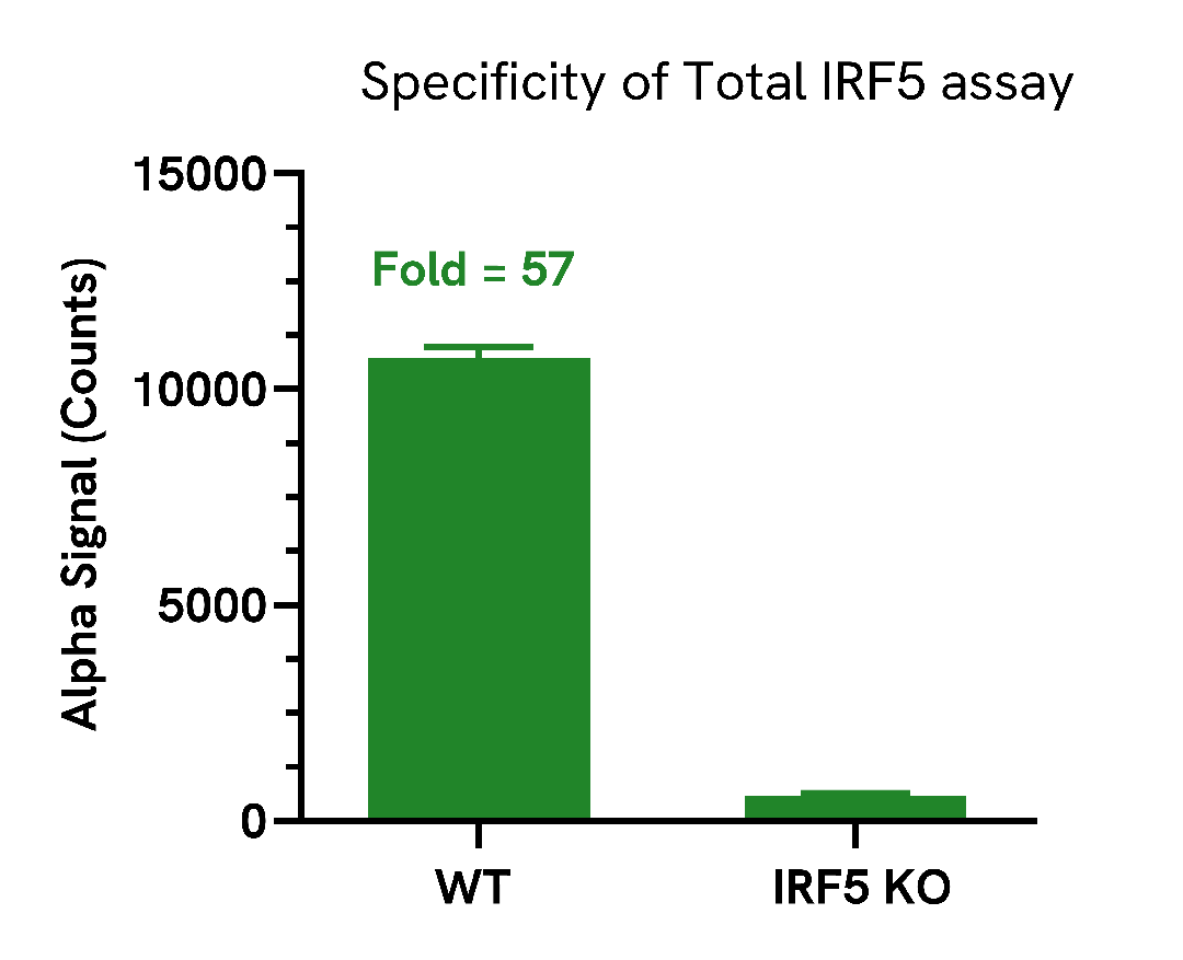
Total IRF5 protein levels were assessed in A549 wild type (WT) and A549 IRF5 knockout (KO) (Abcam ab301006) cells cultured to confluency in T75 flasks at 37°C, 5% CO2.
Each flask was lysed in 2 mL of Lysis Buffer for 10 minutes at RT with shaking (350 rpm). Lysates were then evaluated for Total IRF5 using the AlphaLISA SureFire Biotin Free assay kit.
For the detection step, 10 µL (approximately 10,000 cells) of cell lysate was transferred into a 384-well white OptiPlate, followed by 5 µL Acceptor Mix and incubated for 1 hour at RT. Finally, 5 µL of Donor Mix was added to each well and incubated for 1 hour at RT in the dark. The plate was read on an Envision using standard AlphaLISA settings.
As expected, Total IRF5 protein was detected in WT cells, with very low signal detected in the IRF5 KO cell, demonstrating assay selectivity.
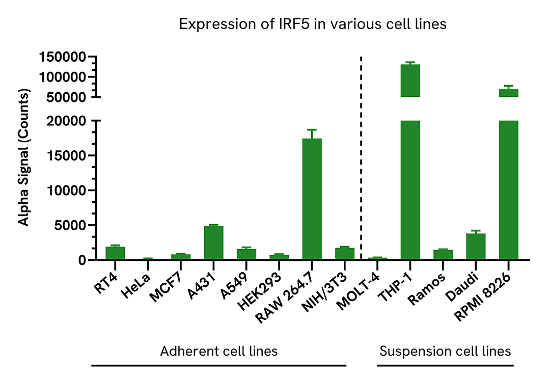
Adherent cell lines were seeded in a 96-well plate (40,000 cells/well) and incubated overnight at 37°C, 5% CO2. Cells were lysed with 200 µL of Lysis Buffer for 10 minutes at RT with shaking (350 rpm).
Suspension cell lines were seeded in a 96-well plate (400,000 cells/well) and lysed with 200 µL of Lysis Buffer for 10 minutes at RT with shaking (350 rpm).
IRF5 levels were evaluated using the AlphaLISA SureFire Biotin Free assay. For the detection step, 10 µL (approximately 2,000 adherent cells and 20,000 suspension cells) of cell lysate was transferred into a 384-well white OptiPlate, followed by 5 µL of Acceptor Mix and incubated for 1 hour at RT. Finally, 5 µL of Donor Mix was then added to each well and incubated for 1 hour at RT in the dark. The plate was read on an Envision using standard AlphaLISA settings.
Expression of IRF5 is varied among human and mouse cell lines. High levels of expression was detected in THP-1, RPMI 8226 and RAW 264.7.
| Application |
Cell Signaling
|
|---|---|
| Automation Compatible |
Yes
|
| Brand |
AlphaLISA SureFire Biotin-Free
|
| Detection Modality |
Alpha
|
| Protocol Time |
2h at RT
|
| Sample Volume |
10 µL
|
| Shipping Conditions |
Shipped in Blue Ice
|
| Target |
IRF5
|
| Target Class |
Phosphoproteins
|
| Target Species |
Human
Mouse
|
| Technology |
Alpha
|
| Therapeutic Area |
Inflammation
|
| Unit Size |
50,000 assay points
|
Loading...


We are here to answer your questions.

