


Revvity Sites Globally
Select your location.
*e-commerce not available for this region.

AlphaLISA SureFire Biotin-Free Human and Mouse Phospho-ERK1/2 (Thr202/Tyr204) Detection Kit, 500 Assay Points

AlphaLISA SureFire Biotin-Free Human and Mouse Phospho-ERK1/2 (Thr202/Tyr204) Detection Kit, 500 Assay Points

AlphaLISA SureFire Biotin Free Phospho Kit Schematic

The AlphaLISA™ SureFire® Biotin-Free Human and Mouse Phospho-ERK1/2 (Thr202/Tyr204) assay is a sandwich immunoassay for quantitative detection of phospho-ERK1/2 (Thr202/Tyr204) in cellular lysates using Alpha Technology.
| Feature | Specification |
|---|---|
| Application | Cell Signaling |
| Protocol Time | 2h at RT |
The AlphaLISA™ SureFire® Biotin-Free Human and Mouse Phospho-ERK1/2 (Thr202/Tyr204) assay is a sandwich immunoassay for quantitative detection of phospho-ERK1/2 (Thr202/Tyr204) in cellular lysates using Alpha Technology.
Product Variants
Unit Size: 100 assay points
Part #:
ASBF-PERK-A-HV
List Price
USD 729.00
Unit Size: 500 assay points
Part #:
ASBF-PERK-A500
List Price
USD 2,463.00
Unit Size: 10,000 assay points
Part #:
ASBF-PERK-A10K
List Price
USD 14,984.00
Unit Size: 50,000 assay points
Part #:
ASBF-PERK-A50K
List Price
USD 48,363.00
For research use only. Not for use in diagnostic procedures. All products to be used in accordance with applicable laws and regulations including without limitation, consumption, and disposal requirements under European REACH regulations (EC 1907/2006).
AlphaLISA SureFire Biotin-Free Human and Mouse Phospho-ERK1/2 (Thr202/Tyr204) Detection Kit, 500 Assay Points
AlphaLISA SureFire Biotin Free Phospho Kit Schematic
AlphaLISA SureFire Biotin-Free Human and Mouse Phospho-ERK1/2 (Thr202/Tyr204) Detection Kit, 500 Assay Points
Product information
Overview
Extracellular signal-regulated kinases 1/2 (ERK1/2) are central components of the MAPK signaling cascade, regulating cell growth, survival, and differentiation. Activated by MEK1/2 through phosphorylation, ERK1/2 phosphorylate diverse cytoplasmic and nuclear targets. ERK1/2 dysregulation is associated with cancer, driving tumor proliferation and resistance to therapy, and with neurodegenerative diseases like Alzheimer's, where it influences amyloid plaque formation.
The AlphaLISA SureFire Biotin-Free Human and Mouse Phospho-ERK1/2 (Thr202/Tyr204) Detection Kit is a sandwich immunoassay for the quantitative detection of phospho-ERK1/2 in cellular lysates, using Alpha Technology.
Formats:
- The HV (high volume) kit contains reagents to run 100 wells in 96-well format, using a 60 μL reaction volume.
- The 500-point kit contains enough reagents to run 500 wells in 384-well format, using a 20 μL reaction volume.
- The 10,000-point kit contains enough reagents to run 10,000 wells in 384-well format, using a 20 μL reaction volume.
- The 50,000-point kit contains enough reagents to run 50,000 wells in 384-well format, using a 20 μL reaction volume.
AlphaLISA SureFire Biotin Free kits are compatible with:
- Cell and tissue lysates
- Antibody modulators
- Biotherapeutic antibodies
- Biotin rich samples
AlphaLISA SureFire Biotin Free kits can be used for:
- Cellular kinase assays
- Receptor activation studies
- High-throughput screening for preclinical studies
Specifications
| Application |
Cell Signaling
|
|---|---|
| Automation Compatible |
Yes
|
| Brand |
AlphaLISA SureFire Biotin-Free
|
| Detection Modality |
Alpha
|
| Protocol Time |
2h at RT
|
| Shipping Conditions |
Shipped in Blue Ice
|
| Target |
ERK1/2
|
| Target Class |
Phosphoproteins
|
| Target Species |
Human
Mouse
|
| Technology |
Alpha
|
| Therapeutic Area |
Neuroscience
Oncology
|
| Unit Size |
500 assay points
|
How it works
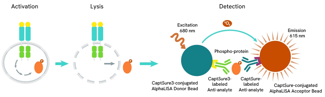
Phospho-AlphaLISA SureFire Biotin Free assay principle
The Phospho-AlphaLISA SureFire Biotin Free assay measures a protein target when phosphorylated at a specific residue.
The assay uses two antibodies which recognize the phospho epitope and a distal epitope on the targeted protein. AlphaLISA assays require two bead types: Acceptor and Donor beads. Acceptor beads are coated with a proprietary CaptSure™ agent to specifically immobilize the assay specific antibody, labeled with a CaptSure tag. Donor beads are coated with a proprietary CaptSure 3 agent to capture one of the detection antibodies, which is labeled with CaptSure 3 tag. In the presence of phosphorylated protein, the two antibodies bring the Donor and Acceptor beads in close proximity whereby the singlet oxygen transfers energy to excite the Acceptor bead, allowing the generation of a luminescent Alpha signal. The amount of light emission is directly proportional to the quantity of phosphoprotein present in the sample.
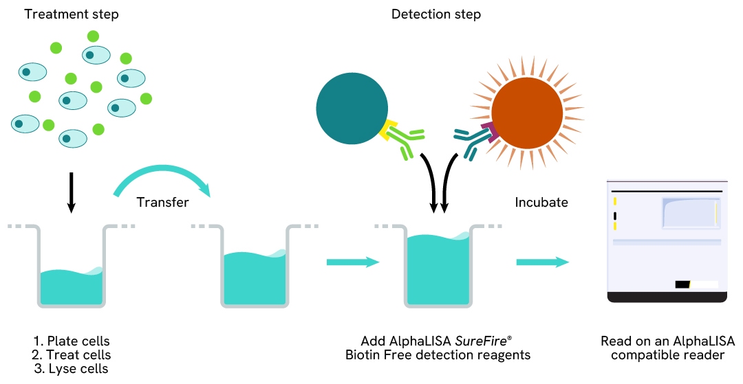
Phospho-AlphaLISA SureFire Biotin Free two-plate assay protocol
The two-plate protocol involves culturing and treating the cells in a 96-well plate before lysis, then transferring lysates into a 384-well Optiplate™ plate before the addition of Phospho-AlphaLISA SureFire Biotin Free detection reagents. This protocol permits the cells viability and confluence to be monitored. In addition, lysates from a single well can be used to measure multiple targets.
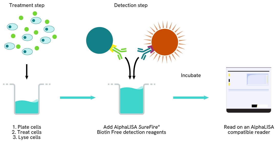
Phospho-AlphaLISA SureFire Biotin Free one-plate assay protocol
Detection of Phosphorylated target protein with AlphaLISA SureFire Biotin Free reagents can be performed in a single plate used for culturing, treatment, and lysis. No washing steps are required. This HTS designed protocol allows for miniaturization while maintaining robust AlphaLISA SureFire quality.
Assay validation
Activation of Phospho ERK1/2 (Thr202/Tyr204) in PMA treated cells
Jurkat cells were seeded in a 96-well plate (200,000 cells/well) in complete RPMI 1640 medium and treated with increasing concentrations of PMA for 15 minutes.
After treatment, the cells were lysed with the addition of 50 µL of 5X Lysis Buffer for 10 minutes at RT with shaking (350 rpm). ERK1/2 Phospho (Thr202/Tyr204) and Total levels were evaluated using respective AlphaLISA SureFire Biotin Free assays. For the detection step, 10 µL of cell lysate (approximately 8,000 cells) was transferred into a 384-well white OptiPlate, followed by 5 µL of Acceptor mix and incubated for 1 hour at RT. Finally, 5 µL of Donor mix was then added to each well and incubated for 1 hour at RT in the dark. The plate was read on an Envision using standard AlphaLISA settings.
As expected, PMA triggered a dose-dependent increase in the levels of Phospho ERK1/2 (Thr202/Tyr204) while Total ERK1/2 levels remained unchanged.

Activation of Phospho ERK1/2 (Thr202/Tyr204) in Anti-CD3 treated cells
Jurkat cells were seeded in a 96-well plate (200,000 cells/well) in serum-free RPMI 1640 medium. The cells were starved for 1 hour and then treated with increasing concentrations of Anti-CD3 antibody for 5 minutes. After treatment, the cells were lysed with the addition of 50 µL of 5X Lysis Buffer for 10 minutes at RT with shaking (350 rpm). ERK1/2 Phospho (Thr202/Tyr204) and Total levels were evaluated using respective AlphaLISA SureFire Biotin Free assays. For the detection step, 10 µL of cell lysate (approximately 8,000 cells) was transferred into a 384-well white OptiPlate, followed by 5 µL of Acceptor mix and incubated for 1 hour at RT. Finally, 5 µL of Donor mix was then added to each well and incubated for 1 hour at RT in the dark. The plate was read on an Envision using standard AlphaLISA settings.
As expected, Anti-CD3 triggered a dose-dependent increase in the levels of Phospho ERK1/2 (Thr202/Tyr204) while Total ERK1/2 levels remained unchanged.

Activation of Phospho ERK1/2 (Thr202/Tyr204) in Anti-IgM treated cells
Ramos cells were seeded in a 96-well plate (200,000 cells/well) in serum free RPMI 1640 medium. The cells were starved for 1 hour and then treated with increasing concentrations of Anti-IgM for 20 minutes. After treatment, the cells were lysed with the addition of 50 µL of 5X Lysis Buffer for 10 minutes at RT with shaking (350 rpm). ERK1/2 Phospho (Thr202/Tyr204) and Total levels were evaluated using respective AlphaLISA SureFire Biotin Free assays. For the detection step, 10 µL of cell lysate (approximately 8,000 cells) was transferred into a 384-well white OptiPlate, followed by 5 µL of Acceptor mix and incubated for 1 hour at RT. Finally, 5 µL of Donor mix was then added to each well and incubated for 1 hour at RT in the dark. The plate was read on an Envision using standard AlphaLISA settings.
Treatment with Anti-IgM triggered a dose-dependent increase in the levels of Phospho ERK1/2 (Thr202/Tyr204) while Total ERK1/2 levels remained unchanged.

Decrease of Phospho ERK1/2 (Thr202/Tyr204) levels in AG1478 treated cells
PC3 cells were seeded in a 96-well plate (40,000 cells/well) in complete Ham’s F-12K (Kaighn’s) medium and incubated overnight at 37°C, 5% CO2. The cells were treated for 2 hours with increasing concentrations of AG1478 and then stimulated with 1 ng/mL of EGF for 30 minutes.
After treatment, the cells were lysed with 200 µL of Lysis Buffer for 10 minutes at RT with shaking (350 rpm). ERK1/2 Phospho (Thr202/Tyr204) and Total levels were evaluated using respective AlphaLISA SureFire Biotin Free assays. For the detection step, 10 µL of cell lysate (approximately 2,000 cells) was transferred into a 384-well white OptiPlate, followed by 5 µL of Acceptor mix and incubated for 1 hour at RT. Finally, 5 µL of Donor mix was then added to each well and incubated for 1 hour at RT in the dark. The plate was read on an Envision using standard AlphaLISA settings.
As expected, AG1478 triggered a dose-dependent decrease in the levels of Phospho ERK (Thr202/Tyr204) while Total ERK1/2 levels remained unchanged.

Resources
Are you looking for resources, click on the resource type to explore further.
Guide
AlphaLISA SureFire Ultra: the ultimate guide for successful experiments
The definitive guide for setting up a successful AlphaLISA SureFire Ultra assay
Several biological processes are regulated by...
Brochure
Alpha SureFire Ultra no-wash immunoassay catalog
Discover Alpha SureFire® Ultra™ assays, the no-wash cellular kinase assays leveraging Revvity's exclusive bead-based technology...
Technical Note
Introducing AlphaLISA SureFire® Biotin Free: A Versatile Immunoassay Platform for Biotin-Rich Samples.
Featuring homogeneous detection of cell-based proteins, this note shows you how to utilize an innovative system optimized for...
Brochure
Species compatibility for HTRF, AlphaLISA SureFire Ultra and Alpha SureFire Ultra Multiplex assays
This document includes detailed tables listing HTRF™, AlphaLISA™ SureFire® Ultra™, and Alpha SureFire® Ultra™ Multiplex assays...


How can we help you?
We are here to answer your questions.

