PhenoVue Microglia Differentiation Staining Kit 1x384
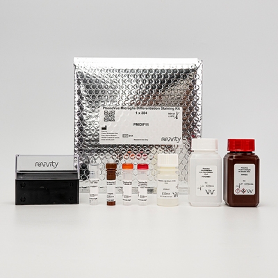
PhenoVue Microglia Differentiation Staining Kit 1x384


Product information
Overview
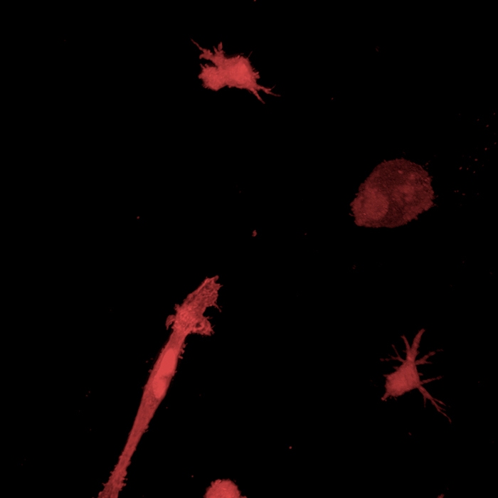
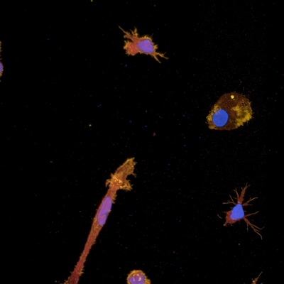
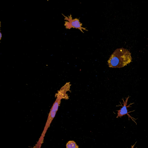
The PhenoVue Microglia Differentiation Staining Kit includes three PhenoVue probes to visualize nuclei, actin and IBA1 protein.
This kit can be used to assess microglia differentiation efficiency from progenitors such as embryonic stem cells or induced pluripotent stem cells (iPSCs), as well as to visualize microglia in brain cell derived co-culture.
The combination of specific, bright and photostable PhenoVue dyes allows a multiplex experiment to be performed with good spectral separation and a significantly reduced spectral overlap.
Each PhenoVue probe has been extensively verified and carefully tested for excellent batch-to-batch reproducibility.
The PhenoVue Microglia Differentiation Staining Kit offers a straightforward and user-friendly protocol to facilitate the experimental procedure.
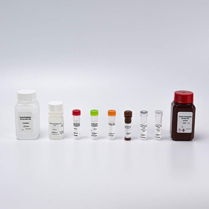
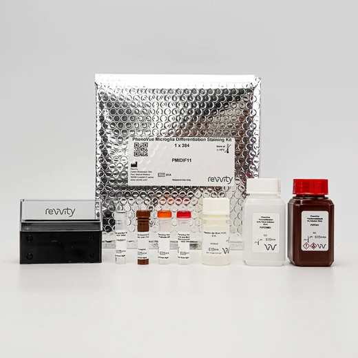
Kit Components / Quantity:
- PhenoVue Hoechst 33342 Nuclear Stain / 1 vial of 70 µL (500x)
- PhenoVue Fluor 555 - Phalloidin / 1 vial (600x)
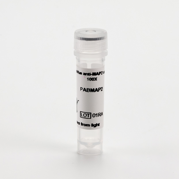
- PhenoVue Anti-IBA1 Antibody (mouse IgG2a) / 1 vial of 100 µL (100x)
- PhenoVue Fluor 647 - Goat Anti-Mouse Antibody Highly Cross-Adsorbed / 1 vial of 100 µL (100x)
- PhenoVue Dye Diluent A (5x) / 1 vial - 8 mL (5x)
- PhenoVue Paraformaldehyde, 4% Solution / 1 bottle of 25 mL
- PhenoVue Permeabilization 0.5% Triton X-100 Solution / 1 bottle of 25mL (5x)
Specifications
| Application |
High Content Imaging
Microscopy
|
|---|---|
| Assay Points |
1 x 384-well microplate
|
| Brand |
PhenoVue™
|
| Detection Modality |
Fluorescence
|
| Quantity |
1 kit
|
| Sample Type |
Fixed samples only
|
| Shipping Conditions |
Shipped in Dry Ice
|
| Storage Conditions |
-16 °C or below, protected from light
|
| Type |
Kit
|
Spectra viewer
Resources
Are you looking for resources, click on the resource type to explore further.
PhenoVue reagents & imaging microplates product list
SDS, COAs, manuals and more
Are you looking for technical documents related to the product? We have categorized them in dedicated sections below. Explore now or request your COA/TDS, SDS, or IFU/manual.
- LanguageEnglishCountryUnited States
- LanguageFrenchCountryFrance
- LanguageGermanCountryGermany


How can we help you?
We are here to answer your questions.


