
Cellaca MX High-throughput Cell Counter
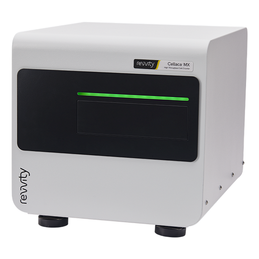
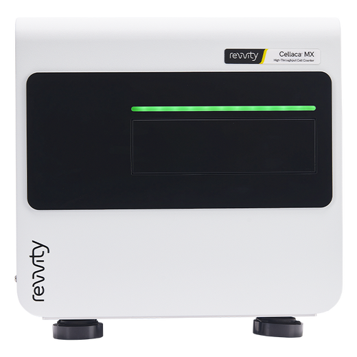
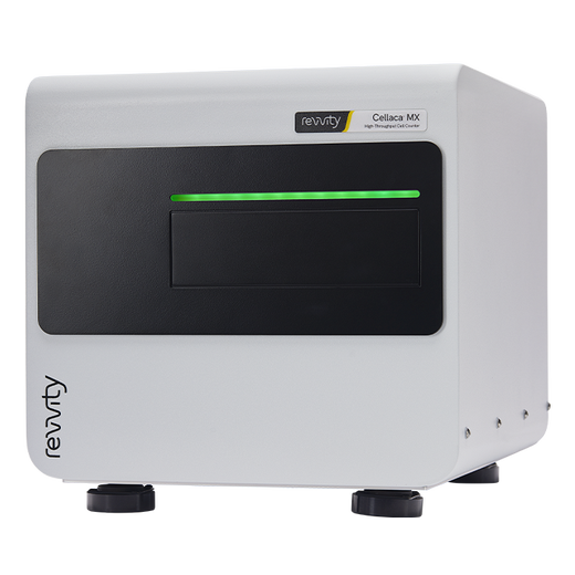
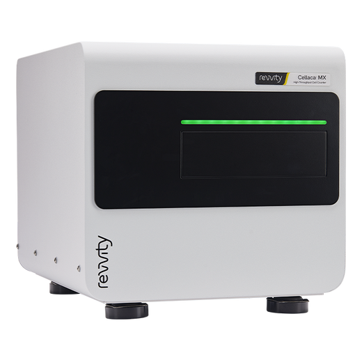


Cellaca MX High-throughput Cell Counter


Cellaca MX High-throughput Cell Counter


The Cellaca™ MX high-throughput cell counter has brightfield and two fluorescent channels. It counts 24 samples with fluorescence in 2.5 minutes or less in a microplate-based format. A 21 CFR Part 11 module is available.
Product information
Overview
Increase your productivity and streamline your workflow with the Cellaca® MX - the high-throughput automated cell counter that changes everything
- Batch samples – run up to 24 samples at a time
- Small sample volume – only 25 µl of cell sample required
- Autofocus – fast autofocus prior to analysis
- Cell based assays – data output at a single cell level enables you to do cell-based assays such as apoptosis, mitochondrial membrane potential, protein expression (including GFP and RFP), and reactive oxygen species (ROS)
- Time course reporting – easily create growth curves with multiple samples at several time points using the customization tools
- Automatic calculation – calculate various data points such as doubling time for individually labeled samples with an easy-to-use interface
- Multiple configurations – choose to add up to five fluorescent filters
- Automation ready – robotic integration ability with optional API
- Configurable reports – add graphs, images, charts, and tables
- Multi-language support – over 7,000 languages available
-
21 CFR Part 11 ready – optional add-on that includes audit trail, user access control, and digital signature
Additional product information
You don’t have to limit your experimental design
Run 24 samples in less than 3 minutes! No more reloading of individual slides or waiting for small batch counters to finish counting. With the Cellaca MX system your experiments are no longer limited by the throughput of your cell counting.
Multiple fluorescent filter options enable you to perform additional cell-based assays in minutes. With the Cell Fitness Panel you can get a quick check of the following cell health parameters:
- Viability
- Vitality
- Apoptosis
- Reactive oxygen species (ROS)

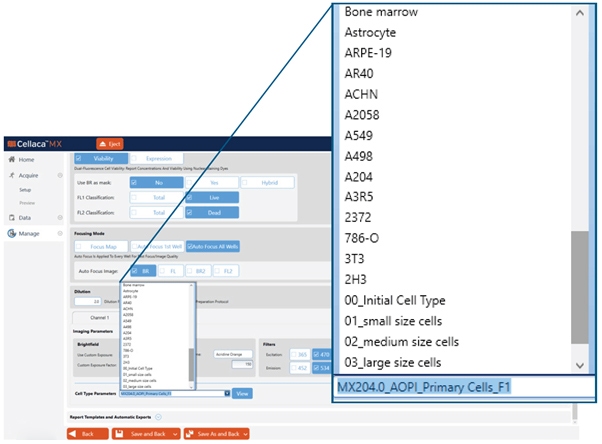
Assay settings and cell type parameters ready to use
With the Cellaca MX cell counter you can build your own assays or choose from the many assays already included in the software. In addition, there are over 200 cell types preloaded with optimized settings. Simply find your assay and cell type in the menu and you are ready to count.
Save time with simultaneous imaging and analysis
As soon as the first well has been acquired you can begin reviewing the images and data. No need to wait until the entire batch has been analyzed before starting to assess the results. Answers are available in seconds, not minutes. The ability to add additional automation with robotic integration can further help increase productivity.
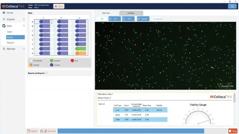
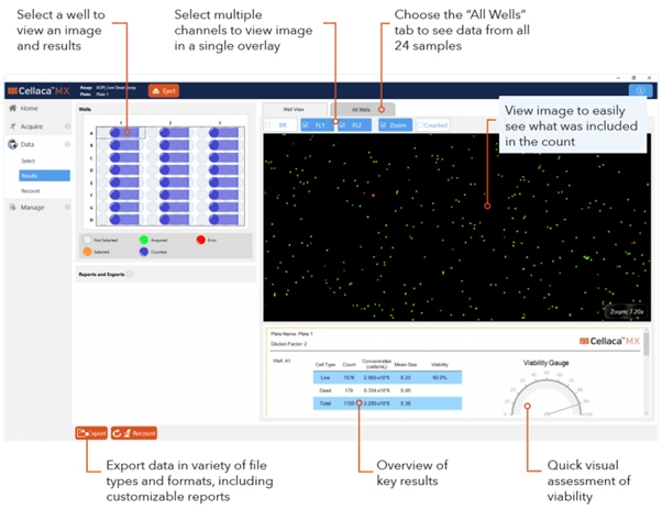
Figure 1. Wells in blue have been captured and analyzed. The green well is in progress of being analyzed. The orange well is next to be analyzed. Well A1 is selected and the AOPI image is displayed on screen (AO is green, PI is red). The report shows the number of live, dead and total cells counted for well A1.
Consistent and Dependable Results
The Cellaca MX system has undergone rigorous testing and verification to ensure reliable performance. In addition, the Cellaca MX counter can alert the operator when the cell count is out of range (too few or too many cells in the sample) and will suggest a corrective course of action (e.g., concentrating or diluting the cell sample and by how much).
Close correlation to hemocytometer
In this study, three tubes of 5-micron polystyrene beads were diluted to a concentration of ~2×10⁶ beads/mL. To determine the concentration within each tube, 40 individual hemocytometer counts were performed for each tube. The Cellaca MX counter was then used to measure the concentration on the same set of tubes. The results show a very close correlation between the hemocytometer counted beads and the Cellaca MX counts.
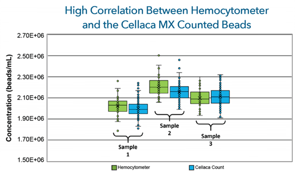
Figure 2. A total of 18 Revvity Counting Plates (432 wells) were used over the 3 tubes of beads and had average CV values of < 4%.
| Hemo CV | Cellaca MX CV | Hemo. Ave | Cellaca MX Ave | % Diff | |
| Tube 1 | 4.46% | 3.69% | 2.03E+06 | E.00E+06 | 1.20% |
| Tube 2 | 4.33% | 3.64% | 2.21E+06 | 2.16E+06 | 2.35% |
| Tube 3 | 4.28% | 3.94% | 2.10E+06 | 2.11E+06 | 0.71% |
Table 2. The measured concentration differences between the Cellaca MX and the hemocytometer bead counts were between 0.7 – 2.35 %.
Low plate-to-plate variability
In a study of 1,992 individually loaded samples (83 plates) by multiple users over multiple weeks a count of ~ 2×10⁶ cells/mL CHO-s cells with Trypan blue yielded a CV less than 6% (figure 3). The cumulative counting time for all 83 plates was 70 minutes.
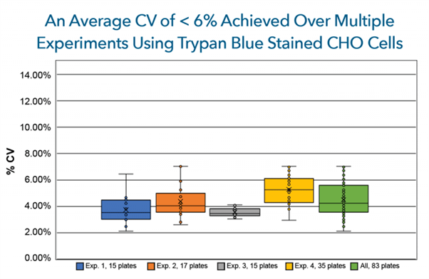
Figure 3. CHO cells stained with Trypan blue were shown to have a CV of 6% or less over 4 independent experiments totaling 83, 24-well plates (1,992 samples).
Consistent Viability Measurement using Fluorescence
Count messy samples more reliably with fluorescence! With the Cellaca MX 2FL and 5FL models, you can reduce red blood cells, platelets and debris from your counts using fluorescent nuclear stains. Acridine orange and propidium iodide are perfect for counting the nucleated cells in samples like PBMCs, patent samples, tumor digests and samples with lots of debris.
The advantage of fluorescent counting for primary cells
The images (Figure 4) demonstrate the advantage of fluorescent counting for primary cells. The bright field image shows the combination of nucleated cells, red blood cells and platelets present in the sample. Only the live and dead nucleated cells are visualized and counted in the green and red fluorescent channels.
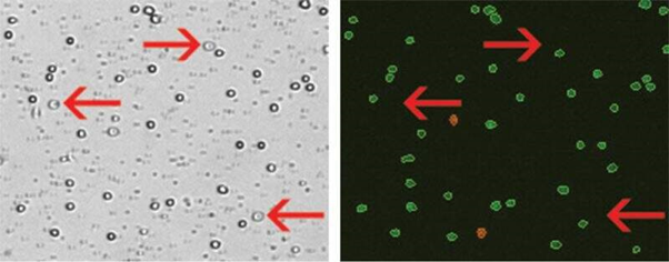
Figure 4. Several red blood cells are indicated in the bright field image (above, left). The red blood cells are not visible in the fluorescent image (above, right) detecting cells stained with nuclear staining dye.
Count Clumpy Cells using Declustering Algorithm
When working with clumpy cells you can apply declustering settings to identify and individually count each cell in your sample to increase the reliability of counts.
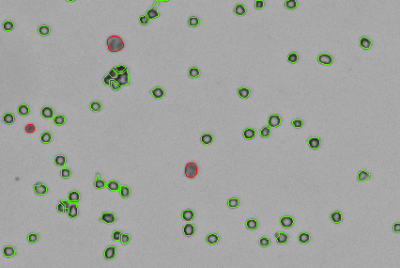
Figure 5. The above image shows cells counted with trypan blue on the Cellaca MX. The software automatically declusters and counts individual live cells (in green outlines) and Trypan blue positive dead cells (in red).
Customizable reports
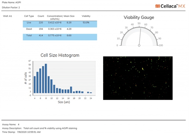
Export reports as CSV, Excel, Word, or PDF files. Use default reports or customize based on the data and images you want included for the assay that was run. Statistical analysis can be included for various parameters. Different languages are available for reporting.
Mix the sample within the plate
Revvity’s proprietary patented* technology allows the user to mix the cell sample and the viability dye or staining reagent in a single well within the plate. The built-in capillary mechanism helps to ensure even distribution across the wells, which provides a completely processed sample set for imaging, counting, and analysis. There is no longer the need to perform these steps in a separate tube.
*U.S. Pat. No. 11,911,764; patents pending: EP3755789A1 and CN201980026197.7
Facilitates 21 CFR Part 11 compliance
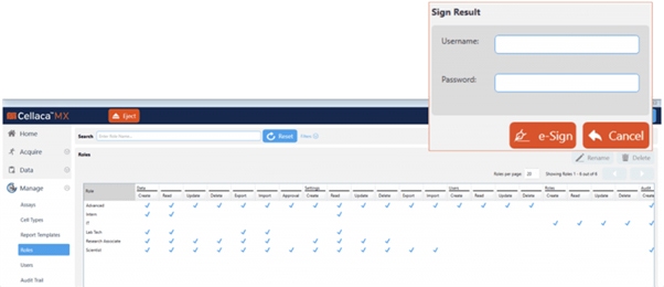
An optional module can be purchased to help facilitate the FDA requirements for 21 CFR Part 11. The additional module comes with the following features:
- User login with passwords
- User-assigned permissions
- Audit trail
- Error log files
- Electronic signatures
Cellaca MX high-throughput cell counter specifications
The Cellaca MX is available in different configurations. Each is field upgradeable to allow your instrument to configure to your experimental needs and quickly drive to deeper cellular insights.
| Cellaca MX BF | Cellaca MX FL2 | Cellaca MX FL5 | |
| Channels | Brightfield | Brightfield, Green, Red | Brightfield, Blue, Green, Red, Far Red |
| Number of Channels | 1 | 4 | 14 |
| Excitation LED | N/A | 470, 527 | 365, 470, 527 and 620 nm |
| Emissions Filters | N/A | 534, 655 | 452, 534, 605, 655 and 692 nm |
| Commonly Used Compatible Dyes | Trypan Blue | Trypan Blue, AO/PI, GFP | Trypan Blue, AO/PI, Hoechst, DAPI, GFP, RFP, CMFDA, Calcein AM, 7AAD, Annexin V |
| Counting Speed Per 24 Samples | 48 seconds | Trypan Blue - 48 seconds Fluorescent - Less than 3 minutes |
Trypan Blue - 48 seconds Fluorescence - Less than 3 minutes |
| Volume (per well) | 25 µL - 100 µL sample volume 50 µL - 200 µL total well volume |
25 µL - 100 µL sample volume 50 µL - 200 µL total well volume |
25 µL - 100 µL sample volume 50 µL - 200 µL total well volume |
| Size/Diameter Range | 5 - 80 µm | 5 - 80 µm | 5 - 80 µm |
| Concentration Range | 1x10⁵ - 1x10⁷ | 1x10⁵ - 1x10⁷ | 1x10⁵ - 1x10⁷ |
| Fluorescence Upgradeable | Yes | Yes (add additional colors) | N/A |
| IQ-OQ Option | Yes | Yes | Yes |
| 21 CFR Part 11 Ready Option | Yes | Yes | Yes |
| Automation Compatible | Yes | Yes | Yes |
| Operating System | Windows 10 | Windows 10 | Windows 10 |
| Dimensions | 13 in x 13 in x 16 in (33 cm x 33 cm x 41 cm) |
13 in x 13 in x 16 in (33 cm x 33 cm x 41 cm) |
13 in x 13 in x 16 in (33 cm x 33 cm x 41 cm) |
| Weight | 40 lbs (18 kg) | 42 lbs (19 kg) | 42 lbs (19 kg) |
Specifications
| Brand |
Cellaca
|
|---|---|
| Unit Size |
1 Each
|
Image gallery


Cellaca MX High-throughput Cell Counter


Cellaca MX High-throughput Cell Counter


Resources
Are you looking for resources, click on the resource type to explore further.
Webinar about integrating synthetic immunology, multiplexing CRISPR, and cell analysis tools to accelerate the development of CAR...
Webinar about streamlining sample preparation for successful single cell sequencing
Webinar about streamlining sample preparation for successful single cell sequencing


How can we help you?
We are here to answer your questions.


