


Total list price:
Log in to view your online price USD 408.57
Select your location.
*e-commerce not available for this region.


 View All


SAVE 10% at Revvity.com or through your punchout with promo code.
SEPT25
Ends Sept. 23 - Terms and conditions apply.
| Feature | Specification |
|---|---|
| Color | Red |
| Filter | Cy5 |
| Organelle and Cell Compartment | Mitochondria |
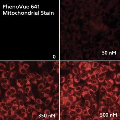
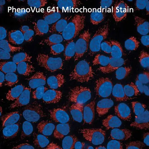
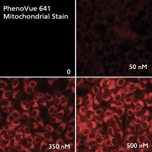
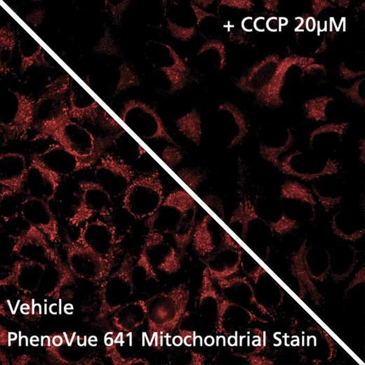
Fluorescent mitochondrial stains are commonly used for visualizing and quantifying mitochondria whose dysfunctions, including impaired biogenesis, dynamics or trafficking, are a hallmark of some human diseases, such as neurodegenerative disorders.
PhenoVue 641 Mitochondrial stain can be used to detect mitochondria in indirect immunofluorescence, immunohistochemistry and flow cytometry, as well as high-content analysis and screening applications.
Typical working concentration: 100 nM (53 ng/mL)
Equivalent number of microplates:
| Color |
Red
|
|---|---|
| Form |
Desiccated
|
| Maximum Emission Wavelength (Emmax) |
662 nm
|
| Maximum Excitation Wavelength (Exmax) |
641 nm
|
| Application |
High Content Imaging
Microscopy
|
|---|---|
| Brand |
PhenoVue™
|
| Detection Modality |
Fluorescence
|
| Filter |
Cy5
|
| Organelle and Cell Compartment |
Mitochondria
|
| Quantity |
20 x 50 µg (92 nmoles)
|
| Sample Type |
Live cells with fixation post staining
|
| Shipping Conditions |
Shipped in Dry Ice
|
| Storage Conditions |
-16 °C or below, protected from light
|
| Type |
Individual reagent
|
Are you looking for resources, click on the resource type to explore further.
This flyer describes Revvity's PhenoVue cellular imaging reagents.
This is a product information sheet for PhenoVue Mitochondrial Stains.
PhenoVue reagents & imaging microplates product list
Are you looking for technical documents related to the product? We have categorized them in dedicated sections below. Explore now or request your COA/TDS, SDS, or IFU/manual.


")


We are here to answer your questions.


