


Operetta CLS Image Gallery
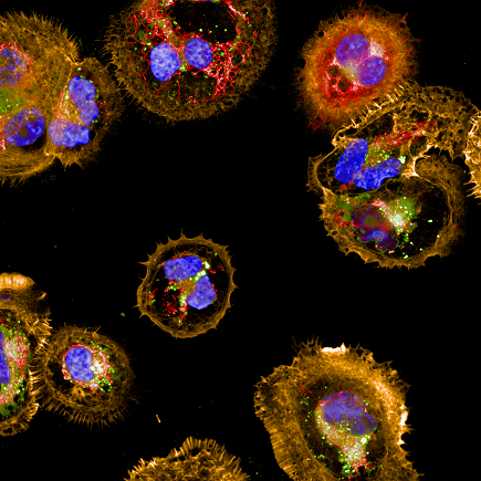
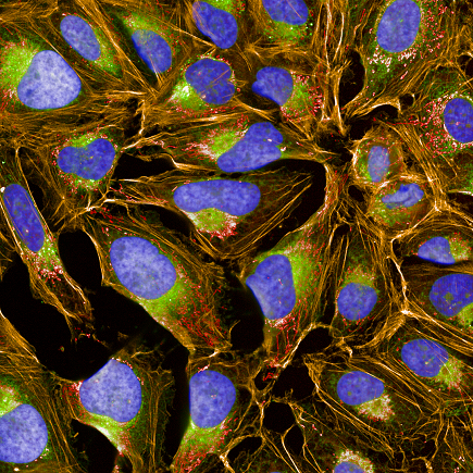
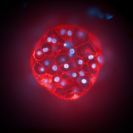
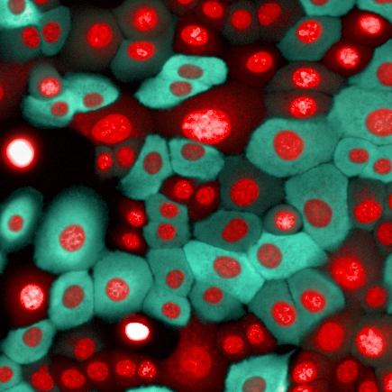
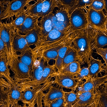
COV434 cells stained with PhenoVue cell painting kit, imaged on Operetta CLS high-content analysis system, 63xW, confocal.
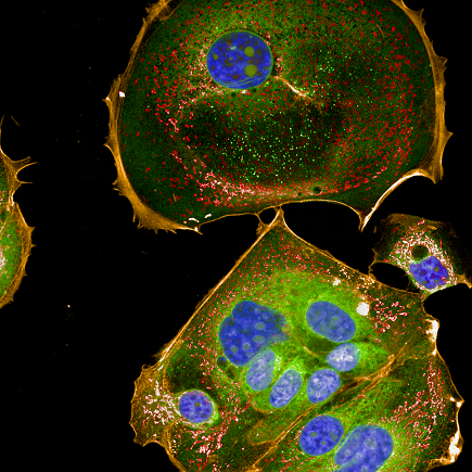
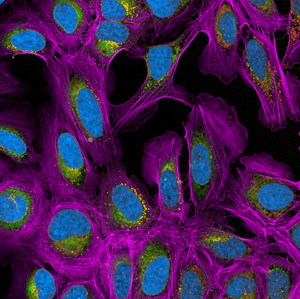
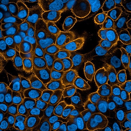
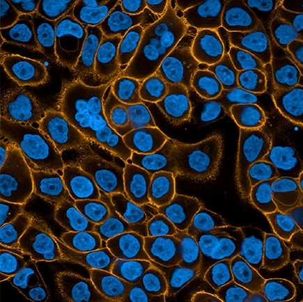
MCF7 cells stained with PhenoVue cell painting kit, imaged on Operetta CLS high-content analysis system, 63xW, confocal.
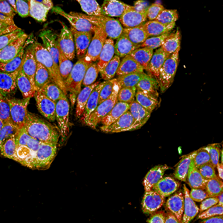
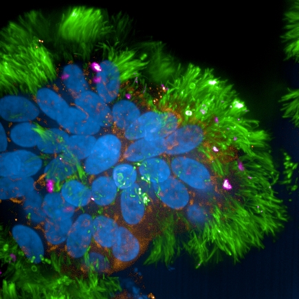
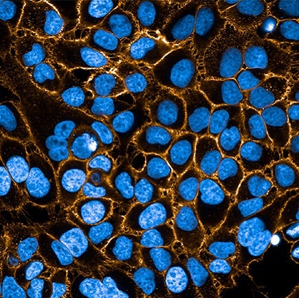
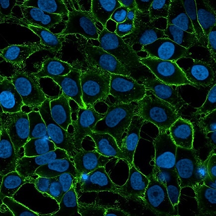
HT-29 cells stained with PhenoVue cell painting kit, imaged on Operetta CLS high-content analysis system, 63xW, confocal.
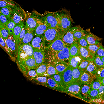
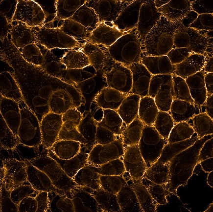
HPAF-II cells stained with PhenoVue cell painting kit, imaged on Operetta CLS high-content analysis system, 63xW, confocal.
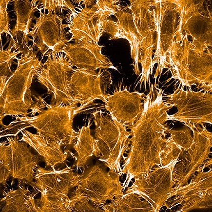
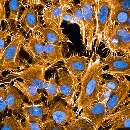
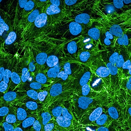
U2OS cells stained with PhenoVue cell painting kit, imaged on Operetta CLS high-content analysis system, 63xW, confocal.
SKOV3 cells stained with PhenoVue cell painting kit, imaged on Operetta CLS high-content analysis system, 63xW, confocal.
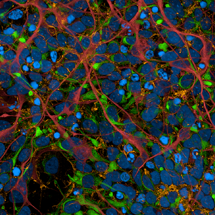
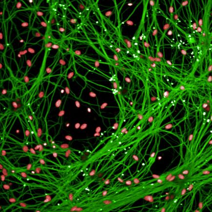
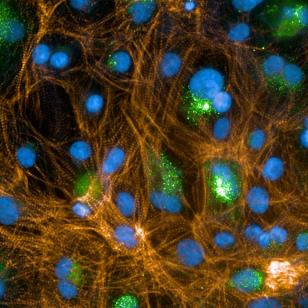
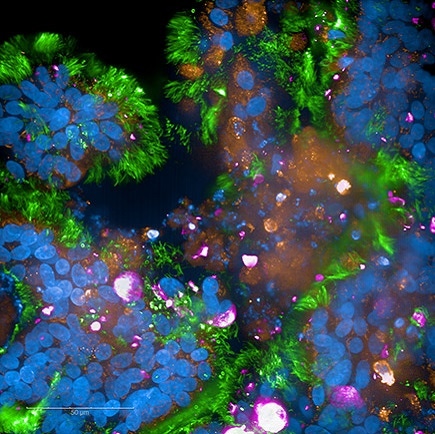
iPSC-derived human cortical neurons stained with PhenoVue neuronal differentiation staining kit, imaged on Operetta CLS high-content analysis system, 63xW, confocal.
U2OS cells 5-plex staining: PhenoVue Hoechst 33342 (blue), Fluor 400LS - Phalloidin (fuchsia), Fluor 488 - Concanavalin A (green), Fluor 555 - WGA (orange), 641 mitochondrial (red). Imaged on Operetta CLS (8 LED) high-content analysis system, 63xW.
SARS-CoV-2-infected apical-out polarized lung organoids. Stained using Hoechst for nuclei (blue), MUC5AC for mucus (orange), phalloidin for tight junctions (pink), and an anti-SARS-CoV-2 antibody (green). Kindly provided by D.Wilfingseder, A.Noureen Institute of Hygiene and Medical Microbiology, Medical University of Innsbruck, Austria
A co-culture of primary dorsal root ganglion neurons and astrocytes. Green = Tuj1, Red = nuclei.
Cells kindly provided by Dr. York Rudhard, Evotec AG, Germany
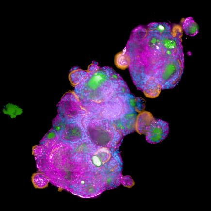
Human Liver Microtissues from InSphero labeled with Hoechst (nuclei, blue) and CellMask™ Deep Red plasma membrane stain.
SARS-CoV-2-infected fully differentiated primary airway epithelia. Stained for SARS-CoV-2 nucleocapsid using a specific antibody (pink), acetylated tubulin for cilia (green), Hoechst for nuclei (blue), and MUC5AC for mucus (orange). Kindly provided by D.Wilfingseder, A.Noureen. Institute of Hygiene and Medical Microbiology, Medical University of Innsbruck, Austria
Human mammary epithelial cells stably expressing the EKAREV biosensor. Nuclei stained with DRAQ5™. Cells imaged using 20xW, confocal. Cell lines kindly provided by Dr. Somponnat Sampattavanich, Department of Pharmacology, Faculty of Medicine Siriraj Hospital, Thailand
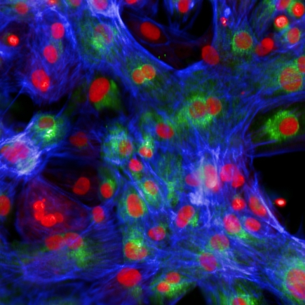
Cor.4U human iPS cell-derived cardiomyocytes labeled with Hoechst (blue), BNP (green) and Rhodamine-Phalloidin (orange) imaged in confocal mode using a 63x water immersion objective. Sample courtesy of Axiogenesis, Cologne, Germany
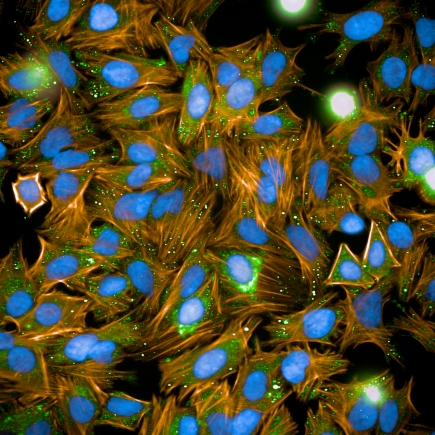
HeLa cells stained with Hoechst (DNA), Alexa Fluor™ 488 labeled anti-tubulin and TRITC-Phalloidin (actin) imaged with a high NA 40x water immersion objective (NA 1.1).
Human cardiomyocytes labeled with the hypertrophy marker proBNP/488, Rhodamine-Phalloidin, Hoechst and HCS CellMask™ Blue, 20x W objective. Cor.4U cardiomyocytes provided by Axiogenesis AG.
A549 non-small cell lung cancer cells treated with EGF to study chemokinesis.
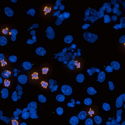
Image showing human cells treated with Nocodazole, labelled with DAPI (nuclei), Alexa Fluor™ 546-labeled antibody for phosphorylated histone H3, Alexa Fluor™ 488-labeled antibody for centrosomes.
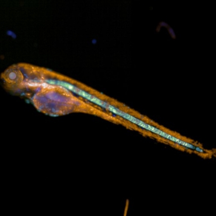
Zebrafish expressing CFP, GFP, YFP and mCherry-tagged proteins. Samples supplied by CCRI, Vienna, Austria
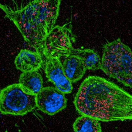
Macrophages with DAPI-labelled nuclei and actin stained with Alexa Fluor™ 488 phalloidin, methicillin-resistant Staphylococcus Aureus labelled with RFP. Image supplied by Dr Penny Tara Mahoney, Vir Biotechnology, San Francisco, CA, USA.

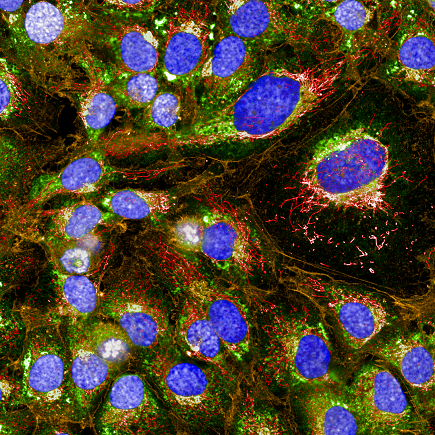
Cells stained with the PhenoVue™ Cell Painting Kit after Berberine chloride treatment, imaged on the Operetta CLS system.
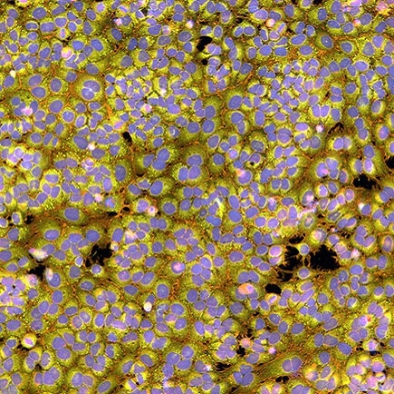
Cells stained with the PhenoVue Cell Painting Kit, imaged on the Operetta CLS system.
SARS-CoV-2-infected fully differentiated primary airway epithelia. Cells were fixed, permeabilized, and stained for SARS-CoV-2 nucleocapsid using a specifc antibody (pink), acetylated tubulin for cilia (green), Hoechst for nuclei (blue), and MUC5AC for mucus (orange). Kindly provided by D.Wilfingseder, A.Noureen. Institute of Hygiene and Medical Microbiology, Medical University of Innsbruck, Austria
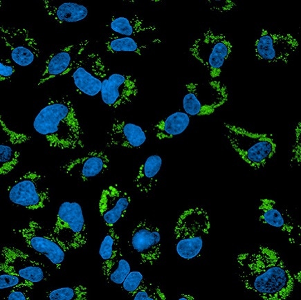
HepG2 cells treated with Oleic Acid-BSA, fixed (PFA), and stained with PhenoVue 493 Lipid Stain (Lipid Droplets, green) + PhenoVue Hoechst 33342 Nuclear Stain (nucleus, blue) (in PBS). Imaged on Operetta CLS.
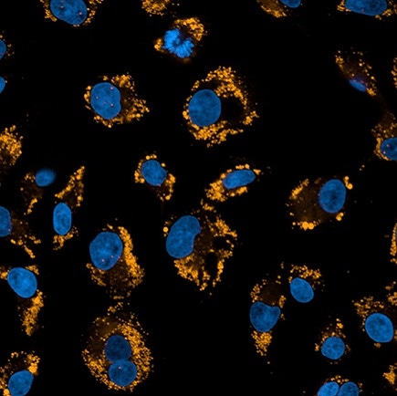
HepG2 cells treated with Oleic Acid-BSA then fixed (PFA) and stained with PhenoVue Nile-Red Lipid Stain (Lipid Droplets, orange) + PhenoVue Hoechst 33342 Nuclear Stain (nucleus, blue). Imaged on Operetta CLS.
A431 cells fixed and permeabilized, stained with Mouse anti EGFR antibody and PhenoVue Fluor 647 - Goat Anti-Mouse Antibody Highly Cross-Adsorbed (EGFR receptor, orange) + PhenoVue Hoechst 33342 (nucleus, blue). Imaged on Operetta CLS.
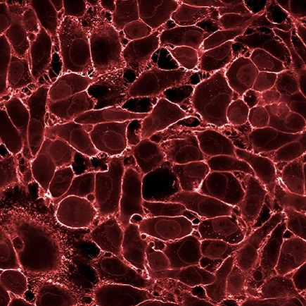
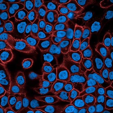
HeLa fixed (PFA) cells stained with PhenoVue Fluor 647 WGA (Plasma Membrane). Imaged on Operetta CLS.
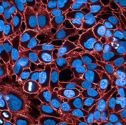
HeLa fixed (PFA) cells stained with PhenoVue Fluor 647 WGA (Plasma Membrane) + PhenoVue Hoechst 33342 Nuclear Stain (nucleus, blue). Imaged on Operetta CLS.
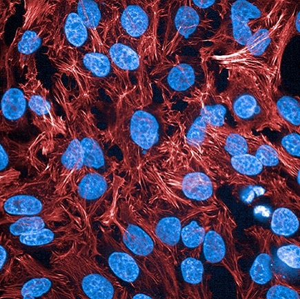
HeLa fixed and permeabilized cells stained with PhenoVue Fluor 647 Phalloidin (actin, red) + PhenoVue Hoechst 33342 Nuclear Stain (nucleus, blue). Imaged on Operetta CLS.

HeLa fixed (PFA) and permeabilized (0.1 Triton X100) cells stained with PhenoVue Fluor 594 WGA (Plasma Membrane + Golgi Apparatus, red) + PhenoVue Hoechst 33342 Nuclear Stain (nucleus, blue). Imaged on Operetta CLS.
A431 cells fixed and permeabilized, stained with Mouse anti EGFR antibody and PhenoVue Fluor 594 - Goat Anti-Mouse Antibody Highly Cross-Adsorbed (EGFR receptor, orange) + PhenoVue Hoechst 33342 (nucleus, blue). Imaged on Operetta CLS.
HeLa fixed (PFA) and permeabilized (0.1% Triton X100) cells stained with PhenoVue Fluor 568 Phalloidin (actin, orange) + PhenoVue Hoechst 33342 Nuclear Stain (nucleus, blue). Imaged on Operetta CLS.
A431 cells fixed and permeabilized, stained with Mouse anti EGFR antibody and PhenoVue Fluor 568 - Goat Anti-Mouse Antibody Highly Cross-Adsorbed (EGFR receptor, orange) + PhenoVue Hoechst 33342 (nucleus, blue). Imaged on Operetta CLS.
HeLa fixed cells stained with PhenoVue Fluor 555 WGA (Plasma Membrane, orange) + PhenoVue Hoechst 33342 Nuclear Stain (nucleus, blue). Imaged on Operetta CLS.
HeLa fixed cells stained with PhenoVue Fluor 555 WGA (Plasma Membrane, orange). Imaged on Operetta CLS.
HeLa fixed (PFA) and permeabilized (0.1% Triton X100) cells stained with PhenoVue Fluor 555 Phalloidin (actin, orange). Imaged on Operetta CLS.
HeLa fixed (PFA) and permeabilized (0.1% Triton X100) cells stained with PhenoVue Fluor 555 Phalloidin (actin, orange) + PhenoVue Hoechst 33342 Nuclear Stain (nucleus, blue). Imaged on Operetta CLS.
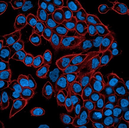
A431 cells fixed and permeabilized, stained with Mouse anti EGFR antibody and PhenoVue Fluor 555 - Goat Anti-Mouse Antibody Highly Cross-Adsorbed (EGFR receptor, orange) + PhenoVue Hoechst 33342 (nucleus, blue). Imaged on Operetta CLS.
HeLa fixed (PFA) cells stained with PhenoVue Fluor 488 WGA (Plasma Membrane, green) + PhenoVue Hoechst 33342 Nuclear Stain (nucleus, blue). Imaged on Operetta CLS.
HeLa fixed and permeabilized cells stained with PhenoVue Fluor 488 Phalloidin (actin, green) + PhenoVue Hoechst 33342 Nuclear Stain (nucleus, blue). Imaged on Operetta CLS.
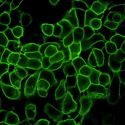
A431 cells fixed and permeabilized, stained with Mouse anti EGFR antibody and PhenoVue Fluor 488 - Goat Anti-Mouse Antibody Highly Cross-Adsorbed (EGFR receptor, green). Imaged on Operetta CLS.
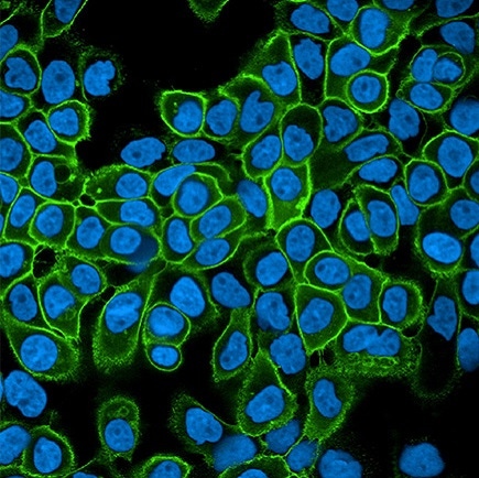
A431 cells fixed and permeabilized, stained with Mouse anti EGFR antibody and PhenoVue Fluor 488 - Goat Anti-Mouse Antibody Highly Cross-Adsorbed (EGFR receptor, green) + PhenoVue Hoechst 33342 (nucleus, blue) (in PBS). Imaged on Operetta CLS.
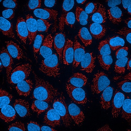
HeLa fixed (PFA) cells stained with PhenoVue 578 Mitochondrial Stain (Mitochondria, red) + PhenoVue Hoechst 33342 Nuclear Stain (nucleus, blue) (in PBS). Imaged on Operetta CLS.
Video shows a 3D rendering of blood vessels in a zebrafish tail recorded with a 20x water immersion lens. Samples supplied by CCRI, Vienna, Austria