
Celigo Image Cytometer


Celigo Image Cytometer


Celigo Image Cytometer


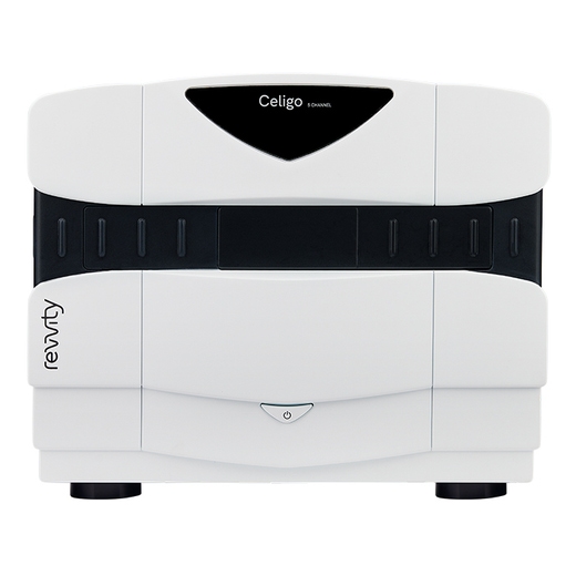
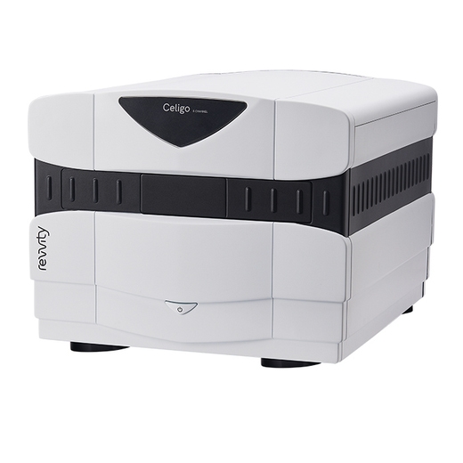
The Celigo™ image cytometer is a microplate-based multichannel brightfield and fluorescent imaging cytometer for 2D and 3D culture using both adherent and suspension cells. A 21 CFR Part 11 module is available.
Product information
Overview
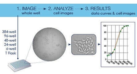
Celigo is a plate-based benchtop brightfield and fluorescent imaging system designed for whole-well live-cell analysis and cell sample characterization. The Celigo system images and analyzes cells in various types of vessels including 6 – 1536 well plates, T25, T75 flasks, 10 cm dishes, and glass slides without disturbing their natural state.
Individual cell level analysis is easily generated, providing cell level insights unlike ELISA or protein-based assays, and at a faster rate than flow cytometry. A broad range of complex cell-based assays have been optimized for the Celigo cytometer:
- Apoptosis
- Cell cycle
- Fluorescent reporters
- Cytotoxicity
- Label-free proliferation
Rapid whole-well imaging and analysis
- Sensitive whole-well imaging ensures accurate cell population analysis
- Remove non-uniform seeding from the equation
- Accurately detect, image, and count every cell in every well
- Specialized scanning mirrors enable fast, high-quality images
Advanced brightfield imaging and four fluorescent channels
- Proprietary algorithm for simultaneous imaging and label-free cell analysis
- Perform real-time multiplex assays with four fluorescent channels
Convenient workflow designed for biologists
-
Multiple object-driven algorithms and a variety of assay-based applications
- Accessible adaptation with easy-to-follow software
- Save customizable experiments as well as default settings
- Built-in gating parameters allow easy data analysis and visualization
- Real-time graphic feedback allows multiple parameters to be measured simultaneously for intuitive analysis
Automation and data management
- Accessible application programming interface (API) for easy automated workflow integration
- Automated microplate handling for kinetic end-point analysis or time-point analysis
Additional product information
Cell counts with whole-well imaging

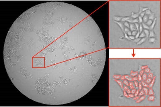
Perform brightfield imaging in combination with four fluorescence channels to provide high speed, fully automated imaging and quantification of suspension and adherent cells, tumor spheroids, iPSC, and cancer stem cell colonies.
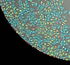
Proprietary optics and scanning system enable fast imaging of the entire well while maintaining consistent illumination and contrast out to the well edge, for identification of cells within each well.
With an intuitive software interface and optional integration into automation platforms, the Celigo provides labs with increased capabilities. Celigo showcases its value from enabling high-throughput screens of drug libraries in early drug discovery through running high-throughput assays for late-stage research.
Brightfield imaging for all well sizes
Excellent optics for enhanced image quality. Improves brightfield optical image quality at the edge of wells and reduces edge optical distortion by using an F-Theta lens for superior well edge-to-edge image contrast.
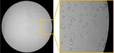
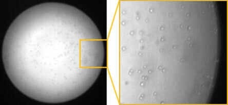
Fast plate scanning for image acquisition and analysis
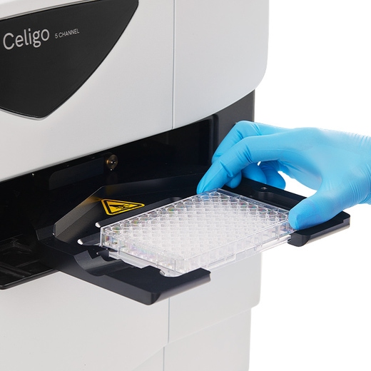
Increase plate-acquisition speed using novel plate scanning technology for fast acquisition. Scan a 384-well plate in brightfield in two minutes or less.
Ability to scan a variety of plate vessels
Scan wells of various sizes using automated image stitching that can view and quantify cells and colonies in vessels up to 6-well plates and 10 cm dishes.
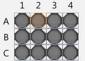
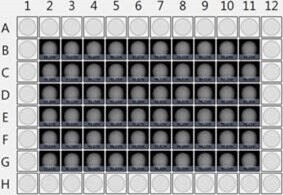
Examples of a 12-well and 96-well plate acquisition using Celigo Imaging Cytometer.
Quantify cells and colonies
Quantify cells and colonies in the well even if they do not grow uniformly across a well.
Measure adherent cells without trypsinization
Analyze your cell sample without trypsinization to help avoid losing cells and look at cells right where they grow over multiple scan times.
Series of customized applications for each assay - ready-to-use
Label-free brightfield cell analysis: Take advantage of label-free brightfield applications to avoid staining cells with toxic dyes or transfecting with fluorescent reporters.
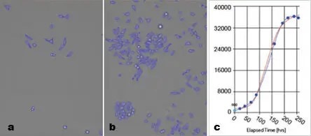
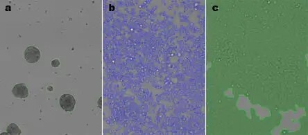
Numerous cell characterization assays: Live cell analysis of images for cell counting, confluence, colonies, and 3D-spheroids.
Monoclonality verification and screening: Detect and evaluate single cell derived colonies over the course of multiple timepoints.
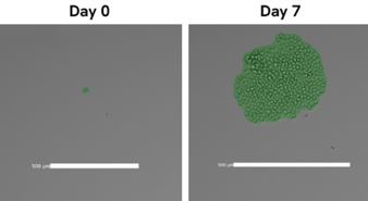
Ability to export brightfield and fluorescent images for publications
Capitalize on the Celigo brightfield and fluorescent image quality to take images of your cells for your records and strengthen your publications.
Easily access and manage data using Celigo network database
Facilitate access to your imaging data using the Celigo network database and connect multiple instruments and satellite workstations to seamlessly acquire and analyze your data from a convenient centralized location.
Easy-to-use preset assay parameters for quick data analysis
Perform Celigo assays using pre-defined parameters that require very few modifications from the researchers.
Simple and intuitive software user interface
Benefit from a simple and intuitive four-step software workflow to run all your assays.
Expand your lab's image analysis & data management capabilities
Storing, managing and sharing large volumes of image data can be a challenge. That's why we've made the Signals Image Artist™ image analysis and management platform from Revvity compatible with Celigo image data for use alongside the Celigo system's own powerful acquisition, visualization and analysis software.
With the addition of the Signals Image Artist platform to your lab, you'll be able to quickly process, store, and share all your image data from the Celigo image cytometer and other major cell imaging systems in a single central location. Its powerful image processing capabilities and ready-made analysis building blocks will provide you with more options for exploring your data and gaining new insights. Benefit from machine learning and artificial intelligence (AI) capabilities to train the software to develop analysis algorithms - and get answers faster. You can even access data and perform analysis remotely via the browser login.
*Signals Image Artist platform is sold separately - it is not sold as part of the Celigo system.
Celigo specifications
| Software |
|
|---|---|
| Illumination/optics |
|
| Plate compatibility |
|
| High-speed imaging |
|
| Weight and dimensions |
|
| Power requirements |
|
| Regularity compliance |
|
Fluorescent channels
| Channel | Excitation | Dichroic | Emission | Typical dyes |
|---|---|---|---|---|
| Blue | 377/50 | 409 | 470/22 | Hoechst, DAPI |
| Green | 483/32 | 506 | 536/40 | FITC, Calcein, GFP,AlexaFluor® 488 |
| Red | 531/40 | 593 | 629/53 | R-PE, PI, Texas Red, AlexaFluor 568 |
| Far-Red | 628/40 | 660 | 688/31 | DRAQ5®, AlexaFluor 647 |
Specifications
| Brand |
Celigo
|
|---|---|
| Unit Size |
1 Each
|
Image gallery


Celigo Image Cytometer


Celigo Image Cytometer


Resources
Are you looking for resources, click on the resource type to explore further.
Cell counting plays a crucial role in the development and manufacturing of cell and gene therapies as well as regenerative...
More than ever, researchers are turning to 3D cell cultures, microtissues and organoids to bridge the gap between 2D cell cultures...
Webinar about 3D cell-based assays in a hypoxic tumor microenvironment
Inhibition of cancer cell proliferation in drug discovery research has not translated well from traditional two dimensional (2D)...
Celigo instrument poster describing A High Throughput Direct Adherent Cell Analysis Method for Cell Cycle and Apoptosis using...
Celigo instrument poster describing A high-throughput 3D tumor spheroid screening method for drug discovery using imaging...


How can we help you?
We are here to answer your questions.


