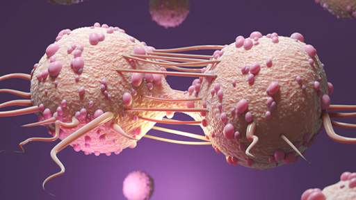
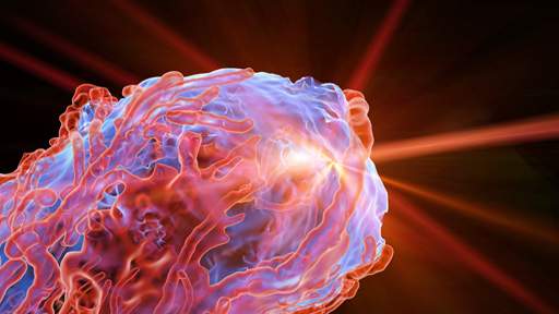
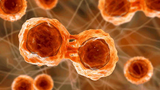
Menu
US

Ultrasound Imaging

Vega™ is a hands-free, automated, high-throughput preclinical ultrasound imaging system that delivers high-resolution 2D and 3D ultrasound images in just a few minutes. This innovative in vivo ultrasound system removes the challenges associated with conventional hand-held systems through the use of automated transducers located under the imaging stage that’s easy to use, requires minimal training and produces more consistent results.

The Vega 3D research ultrasound technology was initially developed and commercialized in 2018 by SonoVol, Inc., a spinout from the Joint Department of Biomedical Engineering at the University of North Carolina at Chapel Hill and North Carolina State University. SonoVol was acquired by Revvity in early 2022. The combined Revvity and SonoVol product offering, including the industry-leading IVIS® optical preclinical imaging platform, provides a comprehensive and powerful solution across ultrasound, optical, and microCT imaging modalities.

For research use only. Not for use in diagnostic procedures.

Request more information
Key features include:
  • Automated transducer positioning and movement allows for a hands-free experience
  • Easy to use with minimal training
  • Enables high-throughput imaging of 3 mice in just a few minutes
  • Whole body field of view
  • Multiple 3D imaging modes: standard B- & M-modes, elastography, and acoustic angiography (CEUS)
  • Small footprint that easily fits on the benchtop

Applications

vega oncology
Oncology
  • Quantify tumor volume
  • Visualize tumor in the context of broader anatomy
  • Deep tissue tumor detection and metastases
  • Visualize tumor microvasculature
radiation research
Liver
  • Measure liver stiffness
  • Quantify steatosis (fat)
  • Evaluate inflammation
  • Visualize liver vascularity
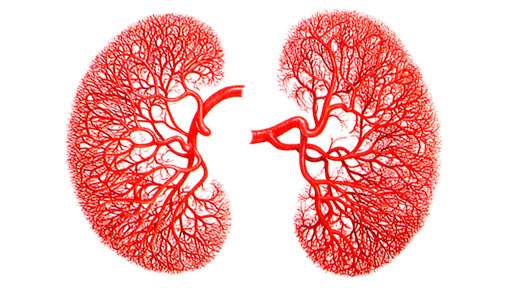
vega kidney vasculature
Kidney
  • Quantify 3D kidney volume
  • Assess changes in kidney perfusion
  • Study kidney microvasculature
vega cardiology
Cardiology
  • Measure cardiac output and ejection fraction
  • Automated heart location
  • Non-gated 4D functional imaging
  • Evaluate drug cardiotoxicity
regenerative medicine
Regenerative medicine
  • Evaluate scaffold vascular patency
  • Track cell seeding over time
radiation research
Radiation research
  • Non-invasively image radiation induced changes to tissue
  • Track volumetric tumor response
  • Evaluate vascular remodeling

Resources