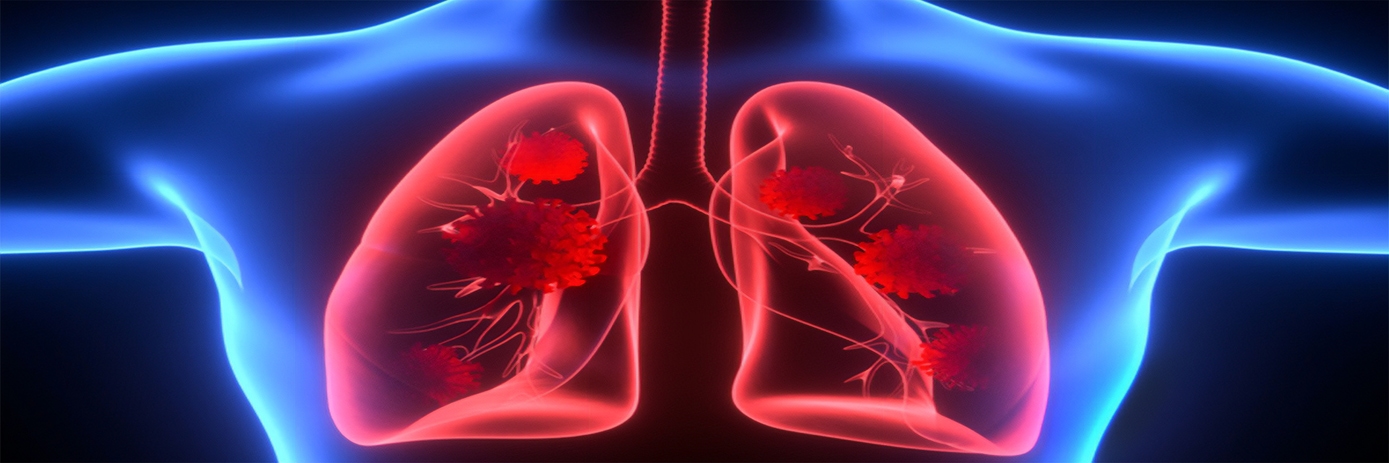
Since the first reported case of COVID-19 in December 2019, researchers around the world have been working to improve their understanding of the novel coronavirus, SARS-CoV-2. Determining the mechanisms by which the virus interacts with the host system is key to developing effective treatments.
A team of researchers in Austria are combining their expertise in three-dimensional (3D) airway cell model development together with high-content analysis to study the first interaction of SARS-CoV-2 with host respiratory tissues.
“Our focus here is on activation of innate immunity by SARS-CoV-2 and whether this activation could be involved in tissue damage and potentially linked to case severity,” said Univ.-Prof. Doris Wilflingseder, from the Institute for Hygiene and Medical Microbiology of the Medical University of Innsbruck (MUI) in Austria.
Exploring interactions in three dimensions
Cell culture models of disease have long been used for basic research and certain clinical in vitro studies. While traditional 2D cell cultures are considered simple, inexpensive, and reproducible, they often poorly mimic in vivo biology.
An important development in cell culture techniques has been the introduction of more physiological relevant model systems, such as 3D cell cultures, spheroids, organoids, and microtissues, that better reflect the living system. Over the last decade, these model systems have become increasingly utilized in certain areas of research and often circumvent the drawbacks of 2D cell cultures and mouse models.
In 2012, Doris’s research group set up an in vitro 3D lung/immune cell model to study the first interactions of Aspergillus fumigatus, an opportunistic fungal pathogen, with upper and lower respiratory tract epithelial and immune cells.1
Over the last few years, the team has worked to further optimize this airway cell model system to work with HIV-1 and, more recently, SARS-CoV-2.
Optimized for success
A key feature unique to the system is that cells are seeded upside down to the bottom side of filters within an animal-free cellulose hydrogel.2
Writing in the journal Cells, the researchers explain that by using this upside-down method, it is possible to transfer the whole insert with cells facing downwards to the glass-bottom dishes for microscopy. “The same samples can be re-used, and live cell imaging can be easily performed without harming the cells,” they said.
At the start of the SARS-CoV-2 pandemic, the group’s system was fully optimized and, together with the working group of Assoc.-Prof. Wilfried Posch at the Institute of Hygiene and Medical Microbiology, Doris’s team are now using their 3D respiratory model to conduct further research on SARS-CoV-2.
She noted that the Operetta™ CLS has become increasingly important for their analysis, providing insights from a single transwell filter of cells. “Not only do you retrieve a lot of information, but you also get fantastic pictures,” she said. “For example, we can visualize the interactions of SARS-CoV-2 with the multilayered epithelia.”
To hear more about the team’s approach and the development of their 3D cell model system, read the full interview with Doris Wilflingseder.
For research use only. Not for use in diagnostic procedures.
References:
- Chandorkar, P., Posch, W., Zaderer, V. et al. (2017) Fast-track development of an in vitro 3D lung/immune cell model to study Aspergillus infections. Sci Rep, 7, 11644. https://doi.org/10.1038/s41598-017-11271-4
- Zaderer, V., Hermann, M., Lass-Flörl, C., Posch, W., Wilflingseder, D. (2019) Turning the World Upside-Down in Cellulose for Improved Culturing and Imaging of Respiratory Challenges within a Human 3D Model. Cells, 8, 1292. https://doi.org/10.3390/cells8101292


