


Monitoring how well a tumor responds to treatment is a critical part of clinical oncology. Response assessment can help diagnose recurrence, determine the efficacy of novel therapeutics in clinical trials, and guide personalized treatment approaches in clinical care.
The gold standard clinical assessment of treatment response is to measure changes in tumor volume over time. Typically, the RECIST (Response Evaluation Criteria in Solid Tumors) guidelines are used to establish whether a tumor disappears, shrinks, stays the same, or gets bigger. However, it often takes weeks or months for changes in tumor volume to occur and RECIST does not assess any other measurements other than tumor size.
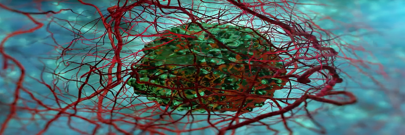
To gain an earlier indication of therapeutic response than RECIST, researchers have explored ways to non-invasively measure molecular or functional changes that occur before tumor shrinkage. One approach is to image the vast network of blood vessels that influence a tumor’s growth, and which play an influential role in treatment response.
Imaging approaches such as contrast-enhanced MRI, perfusion CT, and PET have been widely utilized to visualize blood vessel structure; however, high costs, long imaging times, and radiation risks have limited their clinical application.
An alternative approach is to use a contrast-enhanced ultrasound technique known as acoustic angiography. The benefits of ultrasound imaging are that it is portable, inexpensive, fast, and does not expose patients to ionizing radiation. This makes it an attractive imaging approach compared to MRI, CT, or PET. Furthermore, acoustic angiography enables visualization of the microvascular architecture without a significant contribution from background tissues.
Ultrasound imaging to evaluate treatment response in renal cell carcinoma
A team of researchers recently used ultrasound microvascular imaging in a mouse model of clear-cell renal cell carcinoma (ccRCC) to evaluate whether changes in vascular density indicated treatment response earlier than tumor size assessment.
Anti-angiogenic therapies are often used to treat ccRCC; however, the cancer often becomes resistant shortly after. Early insights into tumor response are therefore critical for effective clinical management or when testing new therapeutic strategies.
For the study, mice bearing ccRCC xenograft tumors were treated with anti-angiogenic and Notch inhibition therapies for four weeks. They were then imaged, and an ultrasound measurement of microvascular density was used to track tumor response to therapy.
The study found that ultrasound measures of microvascular density detected therapeutic response a week earlier than measures of tumor volume. The authors note that while a week may not seem substantial, rodent cancer evolution is much faster than that of humans. “A difference of a week in mice may translate to more clinically relevant time scales in humans,” they said. The authors concluded: “This application of contrast-enhanced ultrasound has important merits as a variety of targeted and immunotherapy agents crowd the treatment landscape of ccRCC.”


