


In the pursuit of novel anti-cancer therapies, researchers need to effectively monitor the efficacy and therapeutic response to promising drug candidates in preclinical models. Standard methods involving bioluminescent reporters, while reliable and highly sensitive, often fall short of providing timely insights by failing to detect metabolic changes prior to overt changes in tumor size. This has led researchers to explore alternative metabolic approaches to gain greater insights into drug-induced responses.
Overcoming limitations of traditional methods
Traditional methods for the preclinical monitoring of anti-cancer drug efficacy primarily rely on physically measuring changes in tumor volume using calipers for subcutaneous tumors or bioluminescence imaging for deeper orthotopic tumors. However, these methods require delayed assessments to detect biological changes that result after prolonged treatment, potentially missing critical early responses.
More recently, the focus has shifted towards measuring alterations in tumor metabolism, which typically occur before physical changes in tumor size become apparent. The gold standard in this approach is 18F-fludeoxyglucose (18F-FDG) positron emission tomography (PET) imaging, a method that measures changes in glucose metabolism.
While 18F-FDG imaging has proven highly effective, its adoption in preclinical research has been limited due to accessibility issues and restrictive safety protocols. These limitations have prompted a search for alternative methodologies that are less complex, more accessible, and cost effective, capable of detecting early therapy-induced metabolic changes in tumors. The goal is to identify methods that are not only effective, but also practical for use in preclinical research.
Exploring other indicators of altered tumor metabolism
Beyond glucose, several other biomarkers are influenced by shifts in tumor metabolism. For instance, metabolically active cancer cells are known to have elevated levels of bombesin and transferrin receptors on their surface. The quantification of these biomarkers offers a potential opportunity to gain insights into altered cellular metabolism and, consequently, evaluate therapeutic response.
To probe this further, Dr. Jen-Chieh Tseng and colleagues set out to study whether fluorescence imaging of bombesin and transferrin receptor expression could detect therapy-induced changes in tumor metabolism in a manner comparable to 18F-FDG PET imaging.1 The researchers utilized two fluorescent imaging probes, IVISense™ Bombesin Receptor 680 (BombesinRSense™) and IVISense Transferrin Receptor 750 (Transferrin-Vivo™), to non-invasively monitor the expression of bombesin and transferrin receptors in response to therapy.
Capturing early therapeutic response through imaging
After establishing a tumor xenograft model, the team administered sorafenib, a clinically approved cancer therapy known for its impact on tumor metabolism. A dose of 40mg/kg was chosen to provide a 2-3 day window of opportunity for early imaging before observable tumor regression in the mice.
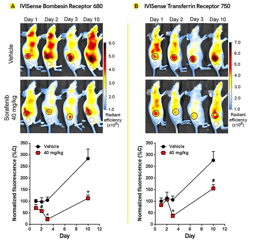
Using PET imaging and the IVIS™ SpectrumCT optical imaging system, the team observed surprisingly comparable results between PET imaging and fluorescence imaging. Figure 1 illustrates the subtle tumor metabolic changes in response to low dose sorafenib, visualized using IVISense Bombesin Receptor and IVISense Transferrin Receptor.
Figure 1: Metabolic changes in HCT116-luc2 tumors were visualized using IVISense Bombesin Receptor 680 and IVISense Transferrin Receptor 750. A: Representative mouse images are shown for 2D fluorescence imaging of IVISense Bombesin Receptor 680 (BombesinRSense 680) tumor uptake, with quantitation of tumor fluorescent signal graphed below. Tumor regions of interest (ROI) are represented by solid line circles, and background control ROI are represented by dotted line circles. B: Representative mice are shown for IVISense Transferrin Receptor 750 (Transferrin-Vivo 750) uptake in tumors, with quantitation of tumor fluorescent signal graphed below. Image credit: Tseng J-C, Narayanan N, Ho G, Groves K, Delaney J, Bao B, et al. (2017) Fluorescence imaging of bombesin and transferrin receptor expression is comparable to 18F-FDG PET in early detection of sorafenib-induced changes in tumor metabolism. PLoS ONE 12(8): e0182689. https://doi.org/10.1371/journal.pone.0182689 licensed under Creative Commons Attribution 4.0 International License.
PET imaging revealed around 55-60% inhibition of tumor uptake of 18F-FDG as early as 2 and 3 days after administering sorafenib. In comparison, the BRS-680 imaging showed 40% and 79% inhibition on days 2 and 3, respectively, while the TfV-750 imaging exhibited 65% inhibition on day 3. Notably, no significant reduction in tumor volume or bioluminescence signal was observed during the initial administration phase.
Writing in PLOS ONE, the researchers explain: “This non-radioactive imaging strategy has the advantage over PET for its capability of simultaneous imaging of multiple biomarkers using probes with different spectral properties.” This capability allowed the team to simultaneously monitor two different biomarkers of interest, with the potential to extend tracking to more than two, leveraging spectral unmixing software that can distinguish the fluorescent channels of each probe.
Final thoughts
The work of Dr. Jen-Chieh Tseng and colleagues showcases the potential of fluorescence imaging as a complementary tool to PET imaging in preclinical research and drug development. By offering correlative insights, but in a more accessible and intuitive format, fluorescence imaging can be considered a powerful approach for monitoring early tumor metabolic responses to targeted therapeutics.
At Revvity, we are committed to supporting advancements in the field of cancer research. Our fluorescent imaging probes are developed through an extensive R&D process, designed to mimic drug-like biodistribution properties, and validated for optimal target delivery and performance to track a broad range of prominent biomarkers.
To help inform your decisions and drive your oncology research forward, we have developed an interactive Selector Guide for IVISense™ fluorescent probes. This resource supports informed decision-making by providing insights into growth, metabolism, protease activity, inflammation, cell death, hypoxia, and other crucial aspects of tumor biology so that you can design oncology studies that maximize your fluorescence readouts.
Reference
- Tseng J-C, Narayanan N, Ho G, Groves K, Delaney J, Bao B, et al. (2017) Fluorescence imaging of bombesin and transferrin receptor expression is comparable to 18F-FDG PET in early detection of sorafenib-induced changes in tumor metabolism. PLoS ONE 12(8): e0182689. https://doi.org/10.1371/journal.pone.0182689
For research use only. Not for use in diagnostic procedures.


