


We’ve all had our blood pressure taken; however, blood pressure measurement isn’t just related to your heart. There is more than one type of hypertension.
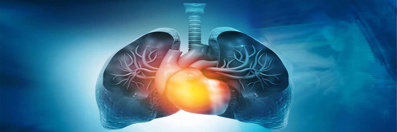
Pulmonary hypertension is caused by high pressure in the arteries that supply blood to your lungs. When the blood vessel walls thicken, blood flow is restricted in a manner similar to a kinked hose, pressure builds up, so the heart must work harder to push the blood through the vessel. This forces the heart to pump more, trying to keep up. Unfortunately, this results in less blood circulating through the lungs causing shortness of breath, dizziness, and fatigue.
Pulmonary hypertension vs. pulmonary arterial hypertension – is there a difference?
The short answer is, yes. Pulmonary Hypertension (PH) is an elevation in the pressure in the arteries of the lungs. PH is associated with diseases such as sleep apnea, fibrosis, COPD, and diseases affecting the left ventricle of the heart; all of which can cause the pressure in pulmonary arteries to rise.
Conversely, Pulmonary Arterial Hypertension (PAH), although a subgroup of PH, is a disease of the blood vessels of the lungs i.e., the vessels have changed causing the increase in pressure. In short, PH is the elevation of pressure caused by another disease, whereas PAH is caused by the blood vessels themselves.
Pulmonary Arterial Hypertension (PAH) is a life-threatening disease where the arteries supplying your lungs are narrowed, blocked, or destroyed. It is characterized by increased pulmonary vascular resistance, elevated pulmonary arterial pressure, and an overload of the right ventricle, which often leads to right heart failure and death. Although considered a rare progressive disease, its overall prevalence is difficult to determine as it is often underdiagnosed.
WHO outlines what causes PAH
What causes PAH is not fully understood. However, there are various risk factors, diseases and substances associated with the disease.
The World Health Organization (WHO) groups PAH into the following categories:
- Idiopathic – can’t be explained by a disease or substance
- Hereditary – Gene BMPR2 (common cause of familial PAH) and ALK1, endoglin, SMAD9, CAV1, KCNK3
- Drugs or toxins (amphetamines, cocaine, fenfluramine-phentermine)
- Association with other diseases (HIV, lupus, rheumatoid arthritis, congenital heart disease)
How is PAH treated?
There are currently no available cures for PAH only treatments to ease its symptoms. These include calcium channel blockers to lower blood pressure, blood thinners, diuretics, and vasodilators, to name a few. Because PAH is currently incurable, more research is needed to understand the pathophysiological mechanisms, identify predictors of prognosis, and development of new therapies.
Right ventricular (RV) function is one of the most important predictors of the disease and is used as a prognostic indicator of RV function in response to PAH therapy. Small animal models such as rodents are ideal for evaluating cardiovascular disease including PAH due to their similarities to human cardiopulmonary physiology potentially enabling translation into the clinic.
In Vivo Imaging’s role
Non-invasive preclinical animal imaging is a well-established tool for understanding disease biology and facilitating drug discovery and development. Various imaging modalities such as MRI, Ultrasound and PET have been used to evaluate cardiac morphology and function in small animal research but high costs, large space requirements, long imaging times, dedicated lab space, complex imaging protocols and repeated radiation exposure (PET) make these less-than-ideal modalities.
Another, and arguably the greatest challenge researchers face when using traditional imaging modalities, is the inability to obtain high resolution images due to motion artifacts from the heart and lung. This makes it difficult to quantify functional changes, for example changes in the right ventricle, and in turn, alterations in disease state and treatment effects over time.
Expanding cardiopulmonary research with MicroCT
MicroCT (micro computed tomography) uses the changes in X-ray attenuation, through objects of varying densities, to reconstruct a 3D image. It provides valuable structural and functional information non-destructively, meaning the subject isn’t altered or destroyed making it ideal for in vivo imaging and long term (longitudinal) studies. Not surprisingly, because microCT uses x-rays to measure variation in density, it is commonly used for skeletal imaging as bones have a high density.
On the other hand, soft, less dense tissue can be more challenging to image using microCT and often requires the use of contrast agents. However, with the development of contrast agents coupled with shorter scan times and ability to produce 3D (3-dimensional) images with high spatial and temporal resolution, microCT has become a powerful tool for non-invasive imaging of micro-architectural structures including lung vasculature.
Also, cardiac, and respiratory motion can be alleviated with ‘gating’ techniques to eliminate motion-induced artifacts and provide accurate quantification over time, making 4D (4-dimensional) imaging possible.
Researchers show how MicroCT was used to assess arterial function in a PAH induced rat model
There is a wealth of published literature showing the usefulness of microCT for quantification of cardiac morphology, stroke volume and cardiac output. Until now, there’s been little to no research published using microCT to assess blood vessel mechanical properties such as vascular distensibility – the ability of vessels to expand or contract – which is an important marker in many disease states including PAH.
However, a team of researchers at the Institute for Lung Health at Justus-Liebig University of Giessen in Germany did just that. By using the Quantum GX low-dose, high resolution microCT imaging system they were able to measure arterial distensibility in a rodent model of PAH. Their research not only showed how fast and easy it was to assess these functional outputs using the Quantum GX microCT (saving both time and money), but more importantly, it showed the value of microCT in strengthening researchers’ knowledge of cardiovascular disease and its potential as a diagnostic and prognostic tool as well as the development of therapeutic strategies for PAH and other cardiovascular disease states.
References
- Kojonazarov et al., Evaluating Systolic and Diastolic Cardiac Function in Rodents Using Microscopic Computed Tomography. Circulation: Cardiovascular Imaging. 11(12) (2018). DOI: 10.1161/CIRCIMAGING.118.007653
- Zelt et al., Micro Computed Tomography in Experimental Pulmonary Arterial Hypertension. Do good things come in small packages? Circulation: Cardiovascular Imaging. 11(12) (2018). DOI: 10.1161/CIRCIMAGING.118.008631


