Cellular imaging allows scientists to study complex cellular dynamics and investigate the cellular basis of diseases such as cancer, metabolic disorders, and infectious diseases. By providing insights into the molecular mechanisms underlying diseases, imaging techniques can aid in the identification of therapeutic targets and contribute to the development of novel diagnostics and therapeutics.
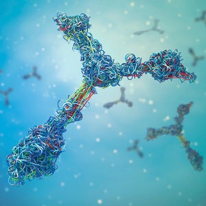
Recent advancements in imaging technologies have revolutionized high-content screening (HCS) workflows, enhancing our ability to track and quantify cellular events. Key to this progress are various imaging reagents and consumables that can be used to prepare and stain samples for analysis. By leveraging tools such as advanced cellular models, gene editing technologies like siRNA and CRISPR, and labeling reagents like fluorescent antibodies and dyes, scientists can modulate and visualize target biology with remarkable clarity.
Here, we explore the significance of imaging reagents in drug discovery research, highlighting how they facilitate the search for innovative therapeutics.
Understanding cellular processes
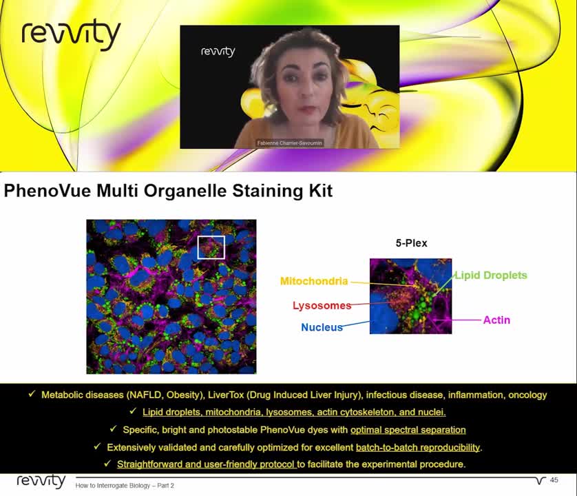
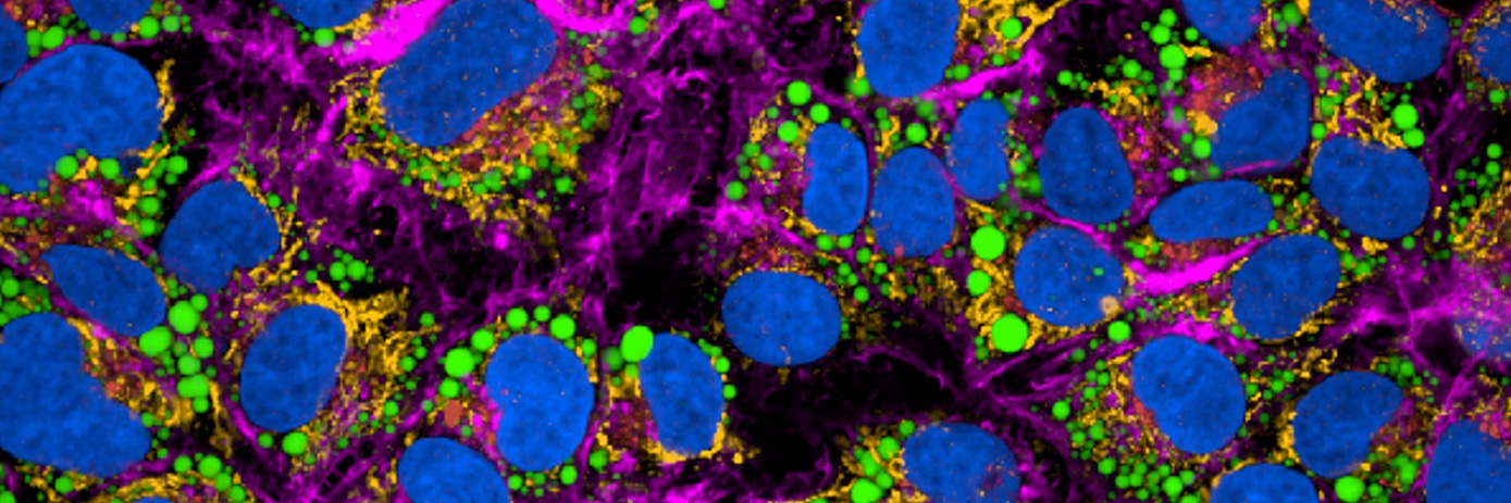
Cellular organelles play crucial roles in various cellular processes, from energy production to genetic regulation. By understanding the dynamics and interactions within cellular compartments, researchers can gain insights into fundamental biological processes and their dysregulation in disease states. Specific organelles can be selectively labeled to facilitate the visualization and tracking of these processes in real time. For instance, labeling mitochondria can reveal insights into cellular respiration and energy production, while nuclear labeling enables the study of DNA replication and gene expression. This can also be achieved through multiplex assays, where multiple organelles and cellular compartments can be labeled and imaged simultaneously.
Investigating cellular events
Imaging reagents can also be used to investigate cellular events such as cell cycle progression, apoptosis, and oxidative stress. Using these tools, morphological changes indicative of apoptosis can be visualized and fluorescent markers that bind to apoptotic proteins can be utilized for detection. Similarly, oxidative stress can be quantified using reactive oxygen species (ROS) probes, providing a deeper understanding of cellular responses to various stimuli.
Advancing disease understanding
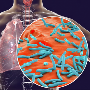
Cellular imaging has become particularly important for investigating disease mechanisms, offering insights into pathological processes. For example, imaging techniques enable the visualization of tumor growth, metastasis, and drug responses at the cellular level, which has become increasingly important in cancer research. Imaging also allows researchers to study essential processes in metabolic disorders, such as lipid metabolism, insulin signaling, and adipocyte function, as well as revealing host-pathogen interactions and immune responses in infectious diseases.
Facilitating drug discovery and development
The integration of imaging reagents into drug discovery pipelines has revolutionized the screening and analysis of potential therapeutic compounds. These reagents facilitate high-throughput screening, allowing for the rapid assessment of numerous compounds against specific cellular targets or pathways. Image-based assays can also provide quantitative data that aid in the identification and optimization of lead compounds, helping to streamline the drug development process. Advancements in imaging technologies have also extended to 3D cultures, enabling researchers to probe complex cellular interactions, such as cell-cell contacts, extracellular matrix remodeling, and drug penetration gradients, within physiologically relevant contexts advancing our understanding of tissue biology and disease mechanisms.
To find out more about our imaging reagents, watch our video below.

Technologies to advance cellular imaging
Key technologies essential for advancing our understanding of cellular structures and functions include imaging microplates, cell painting and staining kits, and hydrogels. At Revvity, we offer a range of solutions covering the entire HCS workflow, from reagents and consumables used for sample preparation and staining to advanced automation and HCS instruments, as well as sophisticated image analysis software.


