


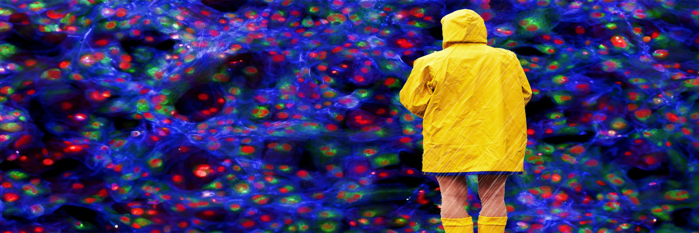

Image-based high-content screening (HCS) systems link cellular imaging with high-throughput analysis enabling the extraction of large amounts of quantitative data from biological systems. In drug discovery, a thorough understanding of the drug target and the drug’s impact on cellular function is needed to identify drug candidates accurately.
The use of image-based HCS systems in drug discovery has expanded in recent years driven by advances in cellular assays, imaging, and analysis technologies that provide greater detail on cellular function and drug mechanism of action. Here, we consider a few of the advances that are increasing the importance of image-based HCS in drug discovery workflows.
Advancements in three-dimensional (3D) cell culture models
3D cell culture models are increasingly used in drug discovery workflows because they more fully reflect the complexities of in vivo environments than traditional 2D cell models. Intercellular interactions and the microenvironment within a tissue can substantially impact cellular responses to a drug. Integrating HCS with 3D cell models allows researchers to gather data from multiple types of live cells within a physiologically relevant context over time. The result is a more accurate and robust understanding of a drug’s impact on cellular structures, events, and processes and the response of both target and non-target cells.
However, despite the promise of 3D cell models, they still come with challenges. 3D cell models present a heterogeneous cell population, which poses analytical challenges for HCS. Advanced detection technologies are being developed to address this challenge and retain the capacity for single-cell profiling within a diverse 3D cell culture. Advanced detection technologies also address the need for high resolution within tissues and cells and maintaining the ability to capture a wide range of signal intensities, both of which can be challenging with 3D cell models.
Multiplexing technologies
Multiplexing enables the simultaneous detection of multiple parameters within a single sample. It is attractive to researchers due to its robust data generation, high efficiency, and cost-effectiveness. In image-based HCS systems, multiplexing leverages different fluorescent dyes, probes, or antibodies for each parameter of interest. A challenge with multiplexing in HCS is the potential for spectral overlap between fluorophores. Advanced imaging techniques are helping overcome this challenge, along with the careful selection of fluorophores with distinct emission spectra, enhancing multiplexing efficacy.
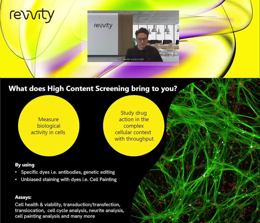
Watch our webinar to learn more about choosing the right physiological model, studying cellular assays with high throughput, and the deeper insights that can be gained with HCS.
Managing the dream data
3D cell models and multiplexing are providing researchers with more data in less time. Couple this with the ability to screen groups of compounds in a single run, and the data can seem overwhelming. Effective data analysis and management tools are crucial for extracting meaningful information from large datasets.
With the increase in data generated by HCS, there is a growing emphasis on developing robust informatics solutions for data storage, management, and analysis. Advanced machine learning algorithms that enhance HCS image acquisition and analysis, including detection and measurement of multiple subcellular structures and processes, are being developed to help automate the identification and classification of complex cellular patterns, enabling more efficient and accurate analysis.


