


Overview
Tumor cells exhibit a different metabolic pattern than normal cells, which includes an increase in glucose uptake (their main source of generating energy). Therefore, cancer cells exhibit an elevated glucose metabolism and up-regulation of glucose transporters (GLUTs) compared to non-tumor cells. IVISense™ 2-DeoxyGlucosone (DG) 750 (formerly XenoLight™ 2-DG) is a fluorescent probe in RediJect™ solution for in vivo targeting of tumors which exhibit an elevated glucose uptake rate in comparison to surrounding tissues. To achieve maximum glucose targeting and enhanced tumor uptake, the probe has been designed to contain four 2-DG molecules per dye molecule.
Products and catalog numbers
| Product | Catalog Number | Ex/Em wavelength (nm) | Validated Experiments | Applications |
|---|---|---|---|---|
| IVISense 2-DG 750 RediJect | 760561 | 750/780 | In vivo | Oncology Inflammation Arthritis |
Using IVISense 2-DG 750 probe in RediJect Solution for in vivo studies
| Product | Route of Injection | Mouse Dose (25 g) | Tissue t 1/2 | Optimal imaging time | Route of Metabolism/ background tissue | IVIS settings |
|---|---|---|---|---|---|---|
| IVISense 2-DG 750 Fluorescent Probe in RediJect Solution | IV | 100 uL | 0.5 h | 3 h | Bladder | IVIS 745/820 |
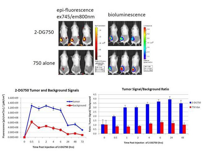
Figure 1: Female C57 BL/6 albino mice were subcutaneously injected with 1 million LL2-luc cells. At 2-3 weeks, tumors were about 100-200 mm3. Mice were intravenously injected with IVISense 2-DG 750 probe (10 nmol/mouse) and imaged with IVIS Spectrum at indicated time points using 745 nm excitation and 800 nm emission filter set. Quantification of the signal from tumor and control background area was plotted on the middle panel graph. Signal to background ratio was shown on the right panel.
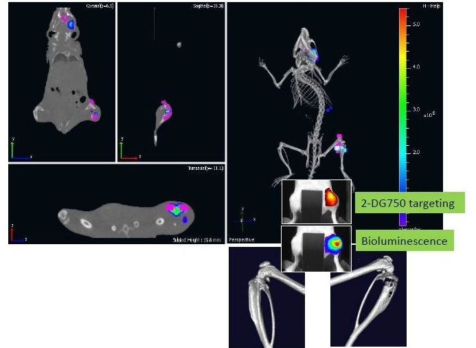
Figure 2: Co-Registered Image Showing Accumulation of IVISense 2-DG 750 at MDA-MB231 Metastasis Sites
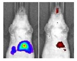
Figure 3: Uptake of IVISense 2-DG 750 by HCT116-luc2 Liver Metastasis. (left) Bioluminescent Signal From Liver Metastases. (right) Epi-Fluorescent Signal From IVISense 2-DG 750 Uptake.
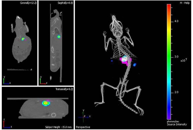
Figure 4: Co-Registration of Bioluminescence (blue) and Fluorescence (magenta) Signals with μCT Image
As indicated above, IVISense 2-DG 750 probe showed specific accumulation to LL-2 tumors shortly after delivery. In addition, specific tumor labeling with the IVISense 2-DG 750 probe has also been demonstrated with Prostate (PC-3M) and Lung (NCI-H460) tumors, further validating the utility of this probe as a generic fluorescence reagent for in vivo tumor targeting.
For research use only. Not for use in diagnostic procedures.