Overview
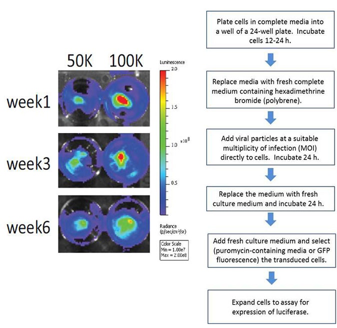
Revvity provides lentiviral particles for creating stably-transfected bioluminescent cells to monitor tumor growth, track primary or stem cells in vivo, and for various other applications using Revvity’s IVIS™ optical imaging platform. Our IVISbrite™ Lentiviral Particles (formerly RediFect™) contain red-shifted firefly luciferase (Luciola italica) or green-shifted Renilla luciferase (Renilla reniformis) transgene and enable efficient transduction of a wide variety of mammalian cell lines including most cancer cell lines, primary, stem and non-dividing cells. Stably transfected bioluminescent cells can be imaged using our luciferin or coelenterazine substrates.

Products and catalog numbers
| Product | Cat. No. | Emission | Notes |
|---|---|---|---|
| IVISbrite Red F-luc-Puromycin | CLS960002 | 620 nm | Puromycin selection marker; in vivo imaging; requires D-luciferin for imaging |
| IVISbrite Red F-luc-GFP | CLS960003 | 620 nm (luciferase) | GFP selection marker; can be used both in vivo (luciferase reporter or dual biolum/fluorescent reporter) and ex vivo (GFP); requires D-luciferin for imaging |
| IVISbrite Green Renilla-Puromycin | CLS960004 | 527 nm | Puromycin selection marker; in vivo imaging; requires coelenterazine for imaging |
Protocols
Creating a stable cell line
- IVISbrite Green Renilla-Puromycin
- IVISbrite Red F-luc-Puromycin
- IVISbrite Red F-luc-GFP
IVISbrite reagent product guiddlines for in vivo and ex vivo applications
- Injection of tumor cells
- Cell preparation and imaging procedure – subcutaneous
- Cell preparation and imaging procedure – intravenous
- Cell preparation and imaging procedure – ex vivo
- Small animal anesthesia protocol
- Imaging Procedure
- Preparation of luciferin for in vitro or in vivo assays using Cat. No. 122796
- IVISbrite D-Luciferin (formerly RediJect D-luciferin) protocol (Firefly luciferase FLuc reporter)
- IVISbrite D-Luciferin Ultra (formerly RediJect D-luciferin Ultra) protocol (Firefly luciferase FLuc reporter)
- Using Coelenterazine H for in vivo imaging (Renilla luciferase RenLuc reporter)
- Determining luciferin kinetic curve for your model
- Intraperitoneal (i.p.) Injection of luciferin
- Intravenous (i.v.) injection of luciferin
FAQs
Q. What is the titer for the IVISbrite lentiviral transduction particles?
A. Each vial contains 1 x 10⁷ units/ml of lentiviral particles resuspended in 200 µl of phosphate buffered saline.
Q. How much virus will I need?
A. The amount of virus necessary to effectively transduce any given cell line must be determined empirically. We suggest titrating the virus to determine the most appropriate amount necessary for your cells. We recommend initial attempts to optimize virus at an MOI range of 10 - 100x. Beyond this range, the lentivirus may become toxic to some cell lines.
Q. Where does the shRNA insert into the genome when delivered with lentivirus?
A. It is commonly believed that lentivirus integrates randomly within the genome. However, some literature suggests that lentivirus is known to insert into active genes.
Q. How many cells can I infect with the amount of lentivirus provided?
A. The amount of cells that can be infected depends on the cell line being used. Primary or other difficult cells may require more lentiviral supernatant. We suggest decreasing the number of cells plated to increase the Multiplicity of Infection (MOI) if necessary.
Q. After selecting puromycin, why are there no surviving cells?
A. The cells you are working with may be sensitive to puromycin. The concentration of puromycin added to the cells may be too high. For each new cell type used, it is recommended that a puromycin titration be performed to determine the lowest concentration of puromycin needed to efficiently select transfected or transduced cells. Also, be sure to wait 24-48 hours post-transduction before applying antibiotics to the cells. Alternatively, a higher MOI or transduction enhancement methods may be required for an effective transduction.
Q. How should a Puromycin titration (kill curve) be set up?
A. Plate 1x10⁴ cells into wells of a 96-well plate with 120 ml fresh media. The next day add 500–10,000 ng/ml of puromycin to selected wells. Examine viability every day. Culture for 2-4 days. The minimum concentration of puromycin that causes complete cell death after 2–4 days should be used for that cell type.
Q. What are the advantages of selection with antibiotics over fluorescent sorting using GFP?
A. Selection of transduced cells can be accomplished using either puromycin or FACS selecting for GFP expressers. That being said, either option is effective. However, for completely pure, transduced cell populations, selection with puromycin is suggested.
Q. What alternatives are available in the event of a polybrene-sensitive cell line?
A. Yes, some cell lines may find polybrene toxic (for example mesenchimal stem cells). Some labs have begun using fibronectin instead.
Q. How many copies/cell can I expect?
A. Some cell lines require a larger dosage of virus (multiplicity of infection). Many researchers aim to only insert a single copy per cell, and this must be determined empirically for each cell line.
Q. Do you sell the corresponding plasmid?
A. No, we are currently not selling the plasmid.
Q. Can I extract the vector directly from the lentiviral particle?
A. No, this is not possible. The vector is used to make the particles in the packaging or producer cells. The viral genome contains only the RNA version of the region found between the 5' and 3' LTRs of pLKO.1 (promoter, RedLuc sequence and puromycin resistance gene or GFP)
Q. Can the lentiviral particles be further propagated in the lab?
A. No, the viral particles cannot be propagated as they are replication incompetent. The particles are made using features of 2nd and 3rd generation lentiviral packaging systems. Genes for replication and structural proteins are absent in the packaged viral genome since these genes are supplied by other plasmids in the packaging cells. The viral genome contains only the region between the 5’ and 3’ LTRs of the plasmid. In addition, the lentiviral vector contains a self-inactivating 3’ LTR that renders it unable to produce infectious virus once it integrates into the host chromosome.
Q. Which packaging system was used to create lentivirus?
A. Third generation packaging system
Q. How serious are lentiviral biosafety concerns? Is there a realistic risk of HIV infection?
A. Viral vector systems have been developed with enhanced safety features. Third generation system consists of:
- The packaging vector, which contains the minimal set of lentiviral genes required to generate the virion structural proteins and packaging functions.
- The vesicular stomatitis virus G-protein (pCMV-VSV-G) envelope vector, which provides the heterologous envelope for pseudotyping
- The transfer vector, which contains the sequence of interest as well as the cis acting sequences necessary for RNA production and packaging. The multi-plasmid approach results in no single plasmid containing all the genes necessary to produce packaged lentivirus.
Resulting particles are replication-incompetent and deletion in the U3 portion of the 3’ LTR eliminates the promoter-enhancer region, further negating the possibility of viral replication. The system has also removed virulence genes which are not necessary for shRNA packaging. These features combined have improved biosafety and handling. It is recommended to use a third generation lentiviral system for its enhanced biosafety features. Though there are no known incidents of third generation systems producing replication competent virus, it is important to monitor for replication competency when performing routine lentiviral packaging. NIH guidelines recommend replication-incompetent lentiviral particles be handled as Risk Group-Level 2 (RGL2). Additional precautions may be required based upon local, state, or country regulations.
Q. Is the RedLuc vector a HIV-based vector and, if so, are there any biosafety issues?
A. The RedLuc vector is a lentiviral (HIV)-based plasmid. The vector is regarded as a biosafety level 2 material and safe to use due to its modified features (deletion of a number of accessory genes implicated in the virulence of HIV, minimal genome of the viral particles, non-replicating and self-inactivation features), making it incapable of producing virus once infected into the host cell. Please consult with your institution’s biosafety officer on specific requirements.
Q. Do I need to modify my lab for work with lentivirus?
A. The NIH recommends preparing your lab for BSL-2 standards when using lentivirus. Please consult with your institution's biosafety officer on specific requirements.
For research use only. Not for use in diagnostic procedures. The information provided above is solely for informational and research purposes only. The information does not constitute medical advice and must not be used or interpreted as such. Consult a qualified veterinarian or researcher for specific guidance or use information. Revvity assumes no liability or responsibility for any injuries, losses, or damages resulting from the use or misuse of the provided information, and Revvity assumes no liability for any outcomes resulting from the use or misuse of any recommendations. The information is provided on an "as is" basis without warranties of any kind. Users are responsible for determining the suitability of any recommendations for the user’s particular research. Any recommendations provided by Revvity should not be considered a substitute for a user’s own professional judgment. Users are solely responsible for complying with all relevant laws, regulations, and institutional animal care and use committee (IACUC) guidelines in their use of the information provided.


